Inflammatory Biomarkers Associated with Symptom Improvement in Chiropractic Neuropathy Patients
July 7, 2025
31 min

Understanding Neuropathy and the Role of Inflammation
Neuropathy, often characterized by pain, numbness, and weakness, stems from complex biological processes involving nerve injury and inflammation. Recent clinical and molecular research has uncovered crucial inflammatory biomarkers that correlate with symptom progression and recovery in various neuropathic conditions. This article explores the emerging scientific evidence connecting inflammatory biomarkers with symptom improvement in chiropractic neuropathy patients, offering insights into their biological roles, clinical implications, and potential mechanisms that bridge inflammation and neurological recovery.
Inflammation as a Key Driver of Neuropathy
 The development of neuropathy often involves complex interactions between nerve injury and inflammatory processes. Inflammatory responses can directly damage nerve structures and also sensitize pain pathways, leading to the characteristic symptoms of neuropathic pain.
Several immune-mediated conditions are major contributors to nerve damage. Autoimmune diseases such as Sjogren’s syndrome, systemic lupus erythematosus (commonly known as lupus), rheumatoid arthritis, and vasculitis are frequently associated with inflammatory peripheral neuropathies. These conditions involve the immune system mistakenly attacking the body's own nerves, resulting in nerve fiber degeneration and dysfunction.
Infections also play a significant role in provoking inflammatory nerve injury. Viruses such as HIV and bacteria like those causing Lyme disease may induce neuroinflammation, further damaging nerves. Certain bacterial toxins, as in botulism, can cause nerve impairment by inflammatory mechanisms. Moreover, autoimmune responses triggered by infections or other conditions, including Guillain-Barré syndrome and chronic inflammatory demyelinating polyneuropathy (CIDP), often include intense immune attacks on peripheral nerves.
Inflammatory markers — including cytokines like IL-1β, IL-6, and TNF-α — are elevated in these conditions and contribute to nerve injury. Systemic inflammation is also observed in metabolic disorders such as diabetes, which is a common cause of diabetic peripheral neuropathy (DPN). Here, low-grade systemic chronic inflammation is linked to nerve damage, with increased levels of pro-inflammatory cytokines correlating with nerve dysfunction.
Understanding the immune components involved in neuropathy has prompted exploration into targeted anti-inflammatory therapies. These include biologics and other immunomodulatory treatments aimed at reducing nerve inflammation and preventing damage.
Overall, inflammation is both a cause and a consequence of nerve injury in various neuropathic conditions, emphasizing the importance of immune regulation in therapeutic strategies.
Overview of Common Inflammatory Biomarkers in Neuropathy

What are common inflammatory biomarkers and their roles?
In the clinical assessment of neuropathy, several inflammatory biomarkers are frequently measured to help understand the underlying inflammatory processes. The most commonly used markers include C-reactive protein (CRP), erythrocyte sedimentation rate (ESR), and plasma viscosity (PV). These markers serve as indicators of systemic inflammation.
CRP is produced by the liver in response to inflammation, and elevated levels suggest the presence of an inflammatory state. ESR measures the rate at which red blood cells settle in a test tube over an hour; faster sedimentation indicates inflammation. Plasma viscosity reflects the thickness of the blood, which can be increased in inflammatory conditions.
While these markers are helpful in detecting general inflammation, they lack specificity—they do not pinpoint the exact cause of inflammation, such as infection, autoimmune activity, or other diseases. Their levels can be influenced by a variety of conditions beyond neuropathy, including infections and chronic inflammatory diseases.
In the context of neuropathy, especially diabetic peripheral neuropathy or inflammatory neuropathies, elevated CRP, ESR, and PV may reflect ongoing systemic inflammation associated with nerve injury. Studies have shown higher levels of these markers in patients with active nerve inflammation, although their levels do not directly correlate with neuropathy severity.
The limitations of these biomarkers stem from their low sensitivity and potential for false positives. Elevated CRP and ESR are common in many ailments, making it challenging to attribute changes solely to neuropathy. Conversely, normal levels do not necessarily exclude neuropathic processes or inflammation.
Therefore, while useful for providing a nonspecific overview of inflammatory status, these biomarkers should be interpreted within the broader clinical picture. They are not definitive diagnostic tools but can serve as part of a comprehensive assessment to monitor disease activity or response to treatment.
| Biomarker | Main Use | Limitations | Additional Notes |
|---|---|---|---|
| CRP | Detects generalized inflammation | Non-specific, elevated in many conditions | Useful for monitoring inflammatory activity |
| ESR | Indicates presence of inflammation | Slow response time, affected by anemia and other factors | Less specific, affected by age and gender |
| Plasma Viscosity | Reflects blood thickness related to inflammation | Less commonly used, less specific | Provides additional inflammatory info |
Role of these biomarkers in neuropathy assessment
While these markers do not diagnose neuropathy directly, their elevation can support evidence of systemic inflammation that may contribute to nerve damage. However, due to their limitations, clinicians rely on a combination of clinical findings, nerve conduction studies, and other specific tests for diagnosis and monitoring.
Specific Inflammatory Biomarkers Linked to Neuropathic Symptomatology

What is the role of cytokines such as IL-6, IL-9, IL-4, TGF-β, and CCL5 in neuropathy?
Cytokines are signaling proteins that play crucial roles in immune responses and inflammation. In the context of nerve injury and repair, cytokines like IL-6, IL-9, IL-4, TGF-β, and CCL5 are particularly important. IL-6 and IL-9 are involved in inflammatory processes that influence nerve healing and pain perception. IL-4 is generally considered anti-inflammatory but has complex interactions with pain pathways. TGF-β and CCL5 are also implicated in systemic inflammation associated with nerve damage.
IL-9, specifically, has been shown to increase at the gene and protein levels following nerve injury, such as in carpal tunnel syndrome. Interestingly, higher levels of IL-9 after surgical intervention were associated with improved pain outcomes, suggesting a role in resolving neuropathic symptoms. Conversely, IL-4 levels tend to be elevated before surgery and positively correlate with pain severity, indicating its involvement during active pain phases.
How do these biomarkers change during nerve injury and recovery?
The levels of these cytokines fluctuate across different stages of nerve injury and healing. For instance, in patients with carpal tunnel syndrome, IL-9 mRNA and protein levels increase after surgical decompression, aligning with symptom improvement. Simultaneously, IL-6 levels tend to decrease with postoperative recovery.
Similarly, TGF-β and CCL5 are elevated in the systemic circulation of patients during active nerve injury, reflecting ongoing inflammation. The decrease in pro-inflammatory mediators like IL-6 post-treatment indicates a dampening of inflammatory responses that may facilitate nerve healing.
Gene expression studies also reveal decreased levels of PTGES2, a gene involved in inflammatory prostaglandin synthesis, during the active phase, which normalizes during recovery. Overall, these dynamic changes highlight the importance of cytokine modulation in nerve repair processes.
Are these biomarkers associated with pain scores and electrodiagnostic severity?
Yes, the levels of certain cytokines directly correlate with pain severity and nerve function assessments. Higher IL-4 protein concentrations are positively associated with increased pain scores and greater electrodiagnostic severity, indicating that IL-4 may be a marker of active nerve injury.
On the other hand, IL-9 gene and protein levels show a negative correlation with pain scores, suggesting that increased IL-9 may help in symptom resolution. This inverse relationship supports the idea that IL-9 could serve as a protective or healing factor rather than a contributor to pain.
In summary, the varying levels of cytokines like IL-6, IL-9, IL-4, TGF-β, and CCL5 during different phases of nerve injury not only reflect the inflammatory environment but also correlate with clinical pain measures. These findings emphasize their potential as biomarkers for monitoring disease activity and recovery in neuropathic conditions.
IL-9: An Emerging Biomarker Associated with Pain Resolution

Increase of IL-9 mRNA and protein after surgical intervention
Recent research on carpal tunnel syndrome (CTS) has highlighted the role of cytokines in nerve injury and recovery. Notably, IL-9 mRNA levels increase following surgical treatment, indicating its potential involvement during the healing process. While IL-9 protein levels also tend to rise, the change has not always reached statistical significance, suggesting variability among patients.
Negative correlation between IL-9 levels and neuropathic pain severity
Interestingly, higher IL-9 levels—both gene expression and protein concentrations—are linked to less severe neuropathic pain symptoms. This negative association suggests that IL-9 may play a part in alleviating pain and promoting nerve repair, thereby contributing to symptom improvement after surgery.
Implications of IL-9 in nerve repair and symptom improvement
The trends observed indicate that IL-9 could be a valuable biomarker for monitoring recovery in patients with nerve injuries such as CTS. Its association with reduced pain severity underscores the possibility that IL-9 not only marks recovery but might actively support nerve regeneration and functional restoration.
Understanding IL-9's precise role could lead to new therapeutic approaches targeting cytokine pathways to enhance nerve healing and pain relief. Ongoing studies are exploring how modulating IL-9 levels might improve patient outcomes, making it a promising candidate for future neuropathic pain management.
Counterbalancing Cytokines: The Case of IL-6 in Neuropathy
How does IL-6 behave during different phases of neuropathic conditions?
Interleukin-6 (IL-6) is a pro-inflammatory cytokine that plays a significant role during the active stages of nerve injury or neuropathy. Studies show that IL-6 levels tend to be elevated during the acute or symptomatic phases, reflecting ongoing inflammation and nerve damage. Elevated IL-6 in serum has been linked with increased pain severity and nerve dysfunction in conditions like carpal tunnel syndrome and diabetic peripheral neuropathy. This cytokine contributes to the inflammatory process, propagating nerve damage if unchecked.
What happens to IL-6 levels as symptoms improve?
Interestingly, after surgical interventions or effective treatments that lead to symptom alleviation, a decrease in IL-6 expression is observed. Research indicates that IL-6 mRNA and protein levels consistently drop as patients recover or report diminished pain. This correlation suggests that dampening IL-6 activity might be associated with the resolution of inflammation and nerve healing. In particular, reductions in IL-6 post-treatment align with improvements in nerve conduction and reduced pain scores, highlighting its potential role as a biomarker for recovery.
Could IL-6 serve as a therapeutic target in neuropathic pain?
Given its prominent involvement in inflammatory pathways, IL-6 presents an attractive target for therapeutic intervention. Strategies that inhibit IL-6 signaling, such as monoclonal antibodies or small-molecule inhibitors, are under exploration for various inflammatory and neurological conditions. Modulating IL-6 activity could potentially reduce nerve inflammation, lessen pain, and promote nerve regeneration. However, more research is needed to determine optimal timing and safety of such targeted treatments, as IL-6 also has roles in immune regulation and repair mechanisms.
| Aspect of IL-6 | Behavior in Neuropathy | Post-treatment Changes | Therapeutic Implications |
|---|---|---|---|
| Levels during active phase | Elevated, correlates with pain severity | Decreased after intervention | Target for anti-inflammatory therapies |
| Role in inflammation | Promotes inflammatory response | Its reduction associates with symptom relief | Modulating IL-6 could help manage inflammation |
| Potential as biomarker | Indicates active nerve injury | Reflects recovery phase | Useful in tracking treatment response |
By understanding the dynamics of IL-6 during nerve injury and recovery, clinicians can better tailor interventions aimed at reducing inflammation and alleviating chronic pain. As research progresses, IL-6 may become a central focus in developing targeted therapies for neuropathic conditions.
Pro-Inflammatory Mediators: TNF-α and Their Role in Diabetic Peripheral Neuropathy

Which biomarkers are associated with neuropathy and its progression?
Research indicates that various biomarkers can reveal the presence and progression of diabetic peripheral neuropathy (DPN). These markers are generally divided into four categories:
- AGE-related molecules: compounds like methyl glyoxal and enzymes such as glyoxalase I, which are linked to the accumulation of advanced glycation end products (AGEs).
- Inflammation-related molecules: including Toll-like receptors, tumor necrosis factor-alpha (TNF-α), miR-146a, and adiponectin. These are involved in inflammatory pathways that may exacerbate nerve damage.
- Nerve damage indicators: specific biomolecules that are directly associated with nerve injury or degeneration.
- Other neuropathic process markers: various mediators that participate in the complex biological processes underlying neuropathy.
Focusing on TNF-α, it emerges as a particularly important cytokine in the context of nerve injury. Elevated levels of TNF-α have been observed in both serum and cerebrospinal fluid of patients with painful diabetic peripheral neuropathy. This cytokine not only characterizes pain in diabetic and other neuropathic conditions but also correlates with nerve function impairments.
TNF-α involvement in painful neuropathies of different origins
TNF-α plays a critical role across different types of neuropathic pain. Its heightened presence is associated with increased inflammatory states that contribute to nerve damage and amplify pain signals. Elevated TNF-α levels have been consistently reported in patients experiencing neuropathic pain, whether diabetic or otherwise. It acts as a central mediator of the inflammatory cascade that leads to nerve degeneration and hypersensitivity.
Association of TNF-α with nerve conduction velocity impairments
High levels of TNF-α are linked to reductions in nerve conduction velocities, especially in nerve fibers like the nervus tibialis. For instance, an inverse relationship has been observed, where increased TNF-α correlates with decreased nerve conduction, indicating worsening nerve function. This association underscores the cytokine's role not only as a marker of inflammation but also as a contributor to nerve deterioration.
Potential of TNF-α as a biomarker for neuropathic pain severity
Given its significant correlation with nerve impairment, TNF-α holds promise as a biomarker for assessing the severity of neuropathic pain. Although current studies suggest its utility, especially in research settings, more validation is necessary before routine clinical application. Nonetheless, measuring TNF-α levels may help in monitoring disease progression and evaluating treatment responses.
| Biomarker Name | Role in Neuropathy | Clinical Correlation | Notes |
|---|---|---|---|
| TNF-α | Inflammatory mediator | Higher levels linked to pain severity | Elevated in serum and CSF, correlates with nerve impairments |
| IL-6 | Pro-inflammatory cytokine | Increased risk of neuropathy | Associated with neuroinflammatory processes |
| miR-146a | MicroRNA involved in inflammation | Potential regulator of cytokine expression | Under investigation |
This evidence collectively supports the importance of systemic inflammatory markers—especially TNF-α—in understanding, diagnosing, and potentially managing diabetic peripheral neuropathy.
TGF-β and CCL5: Indicators of Active Nerve Injury

Elevated serum levels of TGF-β and CCL5 in active neuropathy
During the active phase of carpal tunnel syndrome (CTS), patients exhibit significantly higher levels of systemic inflammatory markers, specifically transforming growth factor-beta (TGF-β) and CCL5, compared to healthy individuals. These molecules are proteins involved in the inflammatory response and play roles in immune regulation.
Their involvement in systemic inflammation during nerve damage
The increase in TGF-β and CCL5 suggests a systemic inflammatory state accompanying nerve injury. These markers are associated with cellular processes such as inflammation, tissue remodeling, and immune cell recruitment, which are integral to nerve damage and repair. Elevated levels indicate that inflammation extends beyond localized nerve compression, contributing to overall systemic responses during nerve injury.
Potential use as markers distinguishing active phases from recovery
Tracking levels of TGF-β and CCL5 can be valuable in clinical practice to differentiate between active nerve injury and recovery stages. During active inflammation, these markers are elevated, whereas they tend to normalize with symptom improvement and nerve healing post-surgery. Their measurement could help clinicians assess disease activity, monitor treatment efficacy, and tailor management strategies for patients with conditions like CTS.
IL-4: Marker of Pain Intensity and Electrodiagnostic Severity

Higher IL-4 protein levels during acute neuropathic pain
Research has shown that IL-4 protein levels are elevated in patients experiencing active neuropathic pain. This cytokine, part of the immune response, appears to be involved in the inflammation processes associated with nerve injury. Elevated IL-4 levels are indicative of the body's immediate response during the acute phase of nerve damage.
Positive correlation with pain scores and nerve conduction studies
Interestingly, IL-4 levels positively correlate with clinical indicators of pain and nerve function. Higher IL-4 concentrations are associated with increased pain severity, as measured by pain scores. They also correlate positively with electrodiagnostic severity, reflecting greater nerve conduction impairment. This suggests that IL-4 not only marks the presence of nerve injury but also reflects the intensity of pain and the extent of nerve damage.
Changes in IL-4 levels post intervention and clinical significance
Post-surgical or therapeutic interventions typically see a decrease in IL-4 levels. This reduction correlates with symptom improvement, indicating that IL-4 could be a useful biomarker for monitoring treatment efficacy. Decreasing IL-4 levels post-treatment may correspond with reduced inflammation and nerve healing, making it a promising target for both diagnosis and tracking recovery progress in neuropathic conditions.
Overall, IL-4 serves as an important marker in neuropathic pain, providing insights into pain severity and nerve health, and potentially guiding treatment strategies.
Molecular Insights from Carpal Tunnel Syndrome Studies
How do systemic cytokine expression profiles in CTS patients change during different phases of the condition?
Research shows that patients with carpal tunnel syndrome (CTS) exhibit elevated levels of systemic inflammatory markers, including cytokines such as IL-9, IL-6, IL-4, TGF-β, and CCL5. During the active phase—when nerve compression and symptoms peak—levels of TGF-β and CCL5 are notably higher compared to healthy controls, indicating ongoing inflammation. Serum IL-9 levels also rise in this phase, and interestingly, IL-9 protein and mRNA levels tend to increase after surgical intervention, correlating with symptom improvement. Conversely, IL-4 levels are higher before surgery and positively relate to pain severity, suggesting its involvement in active pain processes. Elevated inflammatory cytokines such as IL-6 and TGF-β reflect systemic inflammation associated with nerve injury and recovery, making these molecules potential biomarkers for monitoring disease activity and healing.
What does differential gene expression reveal about changes before and after CTS surgery?
Gene expression profiling in CTS patients highlights significant molecular shifts pre- and post-surgery. During the active stage, decreased PTGES2 mRNA indicates suppressed prostaglandin E synthase 2, which may influence inflammation and pain pathways. Post-surgery, a total of 12 genes, including IL-9 and IL-6, show differential expression: IL-9 mRNA levels increase, while IL-6 expression decreases. This pattern suggests that IL-9 might play a role in resolving nerve inflammation and promoting healing, whereas IL-6—known for its pro-inflammatory effects—is involved in active nerve injury. The decrease in IL-4 levels after surgery correlates with reduced pain scores and symptom relief. These molecular changes confirm that cytokine and gene expression modulation are part of the natural course of nerve recovery following decompression surgery.
How do CTS models inform inflammatory biomarker roles in neuropathy?
The human model of CTS provides valuable insights into the inflammatory processes underlying nerve injury and repair. Studies show that cytokines such as IL-9 negatively correlate with pain severity, highlighting their potential role in symptom resolution. Additionally, increased levels of TGF-β and CCL5 observed in patients with CTS suggest systemic inflammation's integral role in nerve damage. The gene expression alterations — especially in cytokines involved in T-cell differentiation and immune responses — underline the immune system's involvement in neuropathic pain. These findings imply that inflammation not only contributes to nerve injury but also influences recovery. Understanding these molecular patterns allows researchers to better grasp how immune responses modulate neuropathy, guiding the development of targeted therapies and biomarkers to improve patient outcomes.
| Aspect | Observations | Implications |
|---|---|---|
| Cytokine levels during CTS | IL-9, IL-6, IL-4, TGF-β, CCL5 elevation | Indicate systemic inflammation and nerve injury phase |
| Post-surgical changes | Increased IL-9, decreased IL-6 and IL-4 | Associated with symptom relief and healing |
| Gene expression | Downregulation of PTGES2; upregulation of IL-9 | Reflect molecular shifts during recovery |
| Model importance | Correlation between cytokines and pain | Guides biomarker development for diagnosis and prognosis |
This molecular understanding emphasizes the significance of inflammatory biomarkers in tracking disease progression and recovery in CTS, with broader implications for neuropathic pain research.
Immune System Dysregulation in Neuropathic Pain Processes

Naïve CD4+ T-cell differentiation cytokine dysregulation
Recent research highlights alterations in cytokine expression involved in naïve CD4+ T-cell differentiation during nerve injury and recovery. These cytokines are critical for guiding immune responses and orchestrating repair processes. Dysregulation of these cytokines can lead to either insufficient nerve regeneration or persistent inflammation, contributing to ongoing neuropathic pain.
Immune involvement in chronic neuropathic conditions
The immune system plays a significant role in chronic neuropathic pain, with systemic inflammatory markers often elevated during different phases of nerve injury. For instance, increased levels of cytokines such as IL-6, IL-1β, and TNF-α are commonly observed in patients with conditions like carpal tunnel syndrome and diabetic peripheral neuropathy. These inflammatory proteins not only reflect ongoing nerve damage but can also exacerbate pain via neuroinflammatory pathways.
Interplay between immune signaling and nerve repair
Immune signaling molecules actively influence nerve repair and regeneration. Cytokines like IL-9, TGF-β, and CCL5 are involved in modulating inflammatory responses and promoting tissue healing. For example, elevated IL-9 after nerve injury has been associated with symptom resolution, suggesting a role in dampening inflammation and facilitating repair. Conversely, decreased expression of certain neuroprotective cytokines can impair nerve regeneration, prolonging pain and dysfunction.
| Cytokine | Role in Neuropathic Pain & Repair | Expression Changes in Studies | Influence on Recovery |
|---|---|---|---|
| IL-9 | Potential anti-inflammatory role during recovery | Increased post-surgery; negatively correlated with pain | Supports nerve healing and reduces pain |
| IL-6 | Involved in inflammation and nerve regeneration | Decreased after surgery; involved in active pain phases | Modulation may aid symptom improvement |
| IL-4 | Associated with immune regulation | Higher pre-surgery levels; positively correlated with pain | Complex role; linked to pain severity |
| TGF-β and CCL5 | Involved in systemic inflammation during nerve injury | Elevated in patients' serum during active stages | May facilitate immune modulation and tissue repair |
Understanding these immune processes provides insights into potential therapeutic targets. Managing cytokine dysregulation could improve nerve healing and alleviate chronic pain, emphasizing the immune system's critical role in neuropathic pain management.
The Dual Role of Systemic Chronic Inflammation in Neuropathy and Pain
How does low-grade systemic inflammation act as a risk factor?
Low-grade systemic inflammation (SCI) is a persistent, non-infective inflammatory state associated with various chronic conditions, including diabetes and obesity. It involves continual release of pro-inflammatory cytokines like IL-6, TNF-α, and IL-1β into the bloodstream.
This ongoing inflammation can damage nerve tissues and disrupt normal nerve function, making individuals more susceptible to neuropathy. Research indicates that higher levels of these cytokines often correlate with nerve impairment and the development of pain symptoms. For example, elevated IL-6 and TNF-α have been linked to increased risk and severity of neuropathic pain.
Is there a connection between systemic inflammation, painful neuropathies, and fibromyalgia?
Yes. Studies have shown that patients with painful peripheral neuropathy and fibromyalgia display heightened levels of inflammatory markers like IL-2, IL-6, and TNF-α in serum and cerebrospinal fluid. These elevated cytokines suggest that systemic inflammation plays a significant role in amplifying pain signals.
Furthermore, long-term low-grade inflammation can not only initiate nerve damage but also sustain chronic pain states. In conditions like fibromyalgia, widespread pain and fatigue are thought to be partly driven by immune dysregulation and neuroinflammation.
How are inflammation and central sensitization linked?
Central sensitization refers to neuroplastic changes within the central nervous system that heighten pain perception, often maintaining chronic pain even when peripheral injury heals. Increasing evidence suggests that systemic inflammation influences brain and spinal cord pathways involved in pain processing.
Inflammatory cytokines like IL-6 and TNF-α can cross the blood-brain barrier, modulating neural circuits and enhancing excitability. This process fosters an environment where pain signals become amplified—a core feature of central sensitization.
Moreover, inflammation can affect brain regions such as the anterior cingulate cortex and thalamus, regions key to pain perception and emotional response. The bidirectional relationship indicates that systemic inflammation can exacerbate central sensitization, and vice versa, establishing a cycle that sustains chronic pain.
| Aspect | Role in Neuropathy and Pain | Explanation |
|---|---|---|
| Risk Factor | Low-grade inflammation predisposes nerves to damage | Chronic cytokine exposure harms nerve tissues, impairing function |
| Association | Elevated inflammatory markers with neuropathic pain | Cytokines are higher in painful neuropathies and fibromyalgia |
| Central Sensitization | Inflammation enhances neural excitability | Cytokines influence brain and spinal cord circuits to increase pain sensitivity |
Understanding this complex interplay helps in developing targeted treatments aiming to reduce systemic inflammation, potentially alleviating nerve damage and chronic pain conditions.
Biomarker Profiles in Diabetic Peripheral Neuropathy

Are inflammatory cytokines useful as early predictors of diabetic peripheral neuropathy (DPN)?
Inflammatory cytokines have attracted attention as potential early markers for DPN. Cytokines like IL-1, IL-6, IL-10, and tumor necrosis factor-alpha (TNF-α) are involved in the inflammatory processes associated with nerve damage in diabetes. Elevated levels of these markers often appear before clinical symptoms emerge, suggesting their potential to serve as early warning signs.
Many studies support the idea that systemic low-grade inflammation correlates with DPN development. Serum levels of pro-inflammatory cytokines tend to rise in patients with early nerve involvement, indicating the transition from metabolic disturbances to nerve injury. Detecting these markers through blood tests could enable earlier diagnosis and intervention, potentially slowing or preventing nerve deterioration.
How do levels of inflammatory biomarkers differ between painful and painless DPN?
Interestingly, while systemic inflammation is generally higher in diabetic individuals with neuropathy, research shows the inflammatory profiles between painful and painless DPN are not markedly distinct.
In fact, patients with painful DPN often display similar or even lower levels of certain cytokines compared to those with painless DPN. For example, cytokines such as TNF-α and IL-6 tend to be elevated in painful neuropathy, reflecting active nerve inflammation, but the overall levels are not highly specific enough to distinguish pain severity reliably.
Moreover, other factors like nerve fiber loss, individual immune responses, and neurochemical changes also influence pain perception, complicating the use of cytokines as straightforward differentiators.
What challenges exist in establishing definitive diagnostic biomarkers for DPN?
Despite promising associations, several hurdles hinder the establishment of reliable biomarkers for DPN. Variability in cytokine levels due to factors like age, comorbidities, and medication use complicates interpretation.
Additionally, cytokine levels can fluctuate over time and are influenced by systemic conditions such as obesity and cardiovascular disease, which are common in diabetics.
Lack of standardized testing methods and consensus on threshold levels further limits clinical application. Many current biomarkers lack specificity for nerve damage related to diabetes, making it difficult to confirm diagnosis solely with blood tests.
Therefore, ongoing research aims to find combinations of biomarkers—possibly integrating cytokines with other inflammatory, neurodegenerative, or metabolic markers—for more accurate and early diagnosis.
| Biomarker | Typical Elevation in DPN | Related Challenges | Potential Role in Diagnosis |
|---|---|---|---|
| IL-6 | Often increased | Fluctuates with systemic inflammation | Early detection when combined with other markers |
| TNF-α | Higher in painful DPN | Not specific to nerve injury | Monitoring disease progression |
| IL-10 | Usually decreased, anti-inflammatory | Variable across individuals | Assessing immune response balance |
| Adipokines (e.g., adiponectin) | Altered levels | Affected by obesity | Supplementary diagnostic data |
Understanding the inflammation-related changes in DPN remains promising, but further large-scale, standardized studies are necessary to translate these biomarkers into routine clinical practice.
Neuroinflammation and Nerve Regeneration Biomarkers
Reduced neuroinflammatory and nerve growth markers in DSPN
Recent studies on diabetic sensorimotor polyneuropathy (DSPN) highlight a surprising reduction in systemic neuroinflammatory and nerve regeneration markers. Patients with DSPN show lower serum levels of cytokines, chemokines, and growth factors that are typically associated with nerve repair and inflammation, such as nerve growth factor (NGF) and certain interleukins. This decrease suggests an impairment in the natural regenerative processes of nerves, potentially contributing to the progression of neuropathy.
Implications for neuropathy pathogenesis beyond inflammation
Traditionally, inflammation has been viewed as a major driver of neuropathic symptoms, especially in diabetic conditions. However, the observed deficits in neuroinflammatory and regenerative biomarkers imply that nerve damage in DSPN might also stem from an insufficient repair response. This shifts some focus from just preventing inflammation to enhancing the body's innate nerve regeneration capabilities, highlighting a dual aspect in the pathogenesis involving both harmful inflammation and inadequate healing.
Potential therapeutic targets based on biomarker deficits
Understanding these biomarker deficits opens new avenues for therapy. Interventions might aim to boost levels of nerve growth factors and cytokines involved in repair, thereby promoting nerve regeneration rather than solely suppressing inflammation. Targeted therapies could include biomolecules designed to enhance neurotrophic support, gene therapies to upregulate regeneration-related genes, or multimodal approaches that combine anti-inflammatory with neurorestorative strategies. Overall, focusing on restoring the balance of neuroinflammatory and regenerative factors could improve outcomes for patients with DSPN.
Proinflammatory Cytokines and Their Impact on Neuropathic Pain
Which proinflammatory cytokines are elevated in painful neuropathy patients?
Research shows that patients suffering from painful peripheral neuropathy often have increased levels of certain proinflammatory cytokines. Notably, cytokines such as interleukin-1 beta (IL-1β), interleukin-2 (IL-2), and tumor necrosis factor-alpha (TNF-α) are found in higher concentrations in these individuals. These cytokines are critical players in immune response and inflammation, which appear to contribute to nerve damage and heightened pain sensations. Elevated levels are typically measured in serum and cerebrospinal fluid, reflecting ongoing inflammatory processes.
How do these cytokines correlate with pain severity and quality of life?
Higher concentrations of IL-1β, IL-2, and TNF-α have been linked to increased pain severity in neuropathy patients. These cytokines can sensitize nerve fibers, making pain signals more intense and persistent. Moreover, their elevated presence is associated with poorer quality of life, as chronic pain impacts daily functioning, emotional well-being, and sleep. For example, studies indicate that patients with higher TNF-α levels tend to report more severe pain and greater psychological distress, underscoring the role of systemic inflammation in overall disease burden.
Can modulating cytokines help in alleviating neuropathic symptoms?
Given the connection between inflammatory cytokines and neuropathic pain, targeting these molecules offers a promising therapeutic avenue. Anti-inflammatory treatments that reduce cytokine levels, such as biologic agents and cytokine inhibitors, are under investigation. By dampening the inflammatory response, it may be possible to decrease nerve sensitization and improve pain outcomes. Some experimental therapies aim to modulate cytokine activity directly, which could result in better symptom control and enhanced quality of life for patients with neuropathy.
| Cytokine | Elevated in Painful Neuropathy | Impact on Pain and Quality of Life | Potential for Therapy |
|---|---|---|---|
| IL-1β | Yes | Increases nerve sensitization, worsens pain | Yes, cytokine inhibitors under study |
| IL-2 | Yes | Promotes inflammation, correlates with pain severity | Potential target for anti-inflammatory drugs |
| TNF-α | Yes | Amplifies nerve damage and pain perception | Drugs that inhibit TNF-α may reduce symptoms |
Understanding the role of these cytokines provides insight into how inflammation drives nerve damage and pain in neuropathic conditions. Targeted therapies aimed at reducing their levels could lead to more effective management strategies, ultimately improving patient outcomes.
Inflammatory Biomarkers in Pediatric and Adolescent Neuropathy
Higher cytokine levels in adolescents with type 1 diabetes and neuropathy
Research shows that adolescents with type 1 diabetes for at least five years exhibit elevated levels of inflammatory cytokines such as interferon-gamma, tumor necrosis factor-alpha (TNF-α), interleukin-10 (IL-10), and soluble urokinase plasminogen activator receptor (suPAR). These increased levels suggest systemic immune activation, which may contribute to nerve damage. Interestingly, these biomarker levels are significantly higher in adolescents with large fiber neuropathy (LFN) compared to those without neuropathy, indicating a possible link between systemic inflammation and nerve injury.
TNF-α association with nerve conduction deficits
Among the cytokines examined, TNF-α stands out for its association with nerve function. Higher TNF-α levels correlate negatively with nerve conduction velocity, especially in the tibial nerve, implying that increased TNF-α may impair nerve signaling. Moreover, elevated TNF-α is associated with higher gastric motility indices in these patients, reflecting broader autonomic and peripheral nerve involvement.
Clinical implications for early diagnosis and intervention
Despite these associations, the studies reveal that inflammatory biomarkers like TNF-α and IL-6 are not reliable screening tools for diabetic neuropathy due to their limited diagnostic accuracy, as indicated by ROC curves with areas under the curve between 0.47 and 0.67. Nonetheless, recognizing elevated inflammatory markers could help identify at-risk adolescents early, prompting interventions aimed at reducing inflammation. Lifestyle modifications, targeted anti-inflammatory therapies, and close neurological monitoring could potentially slow neuropathy progression and improve quality of life.
| Biomarker | Typical Levels in Neuropathy | Correlation with Nerve Function | Diagnostic Utility |
|---|---|---|---|
| TNF-α | Elevated | Lower conduction velocities | Limited, needs further validation |
| IL-6 | Elevated | Associated with neuropathy risk | Limited, with potential in combination biomarkers |
| IL-10 | Increased | Anti-inflammatory, protective | Under investigation |
| suPAR | Elevated | Indicates immune activation | Emerging biomarker |
This ongoing research emphasizes the importance of systemic inflammation in juvenile diabetic neuropathy and highlights the need for further studies to develop reliable biomarkers for early diagnosis and personalized treatment strategies.
Central Sensitization and Its Relationship with Inflammatory Processes
How does central sensitization involve neuroplastic changes that enhance pain sensitivity?
Central sensitization is a process where the central nervous system, particularly the brain and spinal cord, undergoes structural and functional changes that amplify pain signals. These neuroplastic changes include increased excitability of neurons, alterations in neurotransmitter release, and modifications in neural circuitry. As a result, stimuli that were once harmless can become painful, and pain can persist long after the initial injury has healed. This neuroplasticity leads to heightened pain sensitivity, known as hyperalgesia, and can cause non-painful stimuli to be perceived as painful, a phenomenon called allodynia.
In what way does inflammation contribute to the maintenance or perpetuation of central sensitization?
Inflammation plays a significant role in sustaining central sensitization, especially in neuropathic pain conditions. When tissues are injured or inflamed, cytokines such as IL-1β, IL-6, and TNF-α are released, activating immune cells and sensitizing peripheral nerves. These inflammatory mediators can also cross the blood-brain barrier or be produced within the central nervous system, further activating spinal and brain neurons involved in pain processing. Chronic low-grade inflammation, often seen in conditions like diabetic neuropathy or multiple sclerosis, enhances neuroplastic changes by promoting excitatory signaling and inhibiting inhibitory pathways, thus maintaining the heightened pain state.
What are the clinical implications of understanding the link between inflammation and central sensitization?
Recognizing the connection between inflammation and central sensitization has important treatment implications. It suggests that therapies aimed at reducing systemic or local inflammation could help reverse or mitigate central sensitization. This includes anti-inflammatory medications, lifestyle interventions to decrease chronic low-grade inflammation, and non-pharmacologic approaches like stress management and diet modifications. Furthermore, understanding this link underscores the importance of comprehensive pain management strategies that combine immune modulation with therapies targeting neural plasticity, which can improve outcomes in chronic pain conditions like fibromyalgia, neuropathic pain, and complex regional pain syndrome.
| Aspect | Effect | Clinical Relevance |
|---|---|---|
| Neuroplastic changes | Increased excitability, circuit rewiring | Amplifies pain perception, sustains chronic pain |
| Inflammatory mediators | Cytokine release, immune activation | Maintains sensitized neural state |
| Treatment approaches | Anti-inflammatory, neuroplasticity-modulating | Targeting inflammation can reduce central sensitization |
Understanding these processes highlights the importance of a multidimensional approach to managing complex, chronic pain conditions driven by central sensitization.
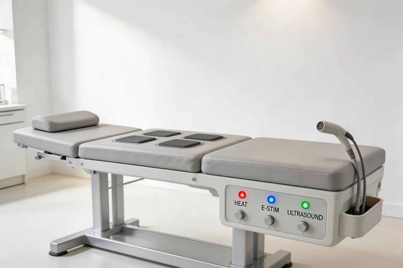
Non-Invasive, Non-Pharmacological Therapies Targeting Inflammation
TENS, heat therapy, massage, light therapy, and neurostimulation
Various non-pharmacological interventions such as Transcutaneous Electrical Nerve Stimulation (TENS), heat therapy, massage, light therapy, and neurostimulation are increasingly used to manage chronic pain, especially where inflammation plays a central role. TENS activates large Aβ fibers, inhibiting pain signals at the dorsal horn. Heat and cold therapies influence neural plasticity, enhancing endogenous opioid release and reducing neuroinflammation. Massage not only relaxes muscles but also promotes the release of natural painkillers and modulates inflammatory cytokines.
Light therapy, including low-level laser therapy, improves mitochondrial function and reduces neuroinflammation, which can decrease pain sensitivity. Neurostimulation techniques like pulsed radiofrequency and vagus nerve stimulation target neural circuits involved in pain and inflammation, helping modulate abnormal immune responses.
Effects on inflammatory mediators and central sensitization
These therapies impact neurochemical pathways involved in inflammation and central sensitization. They reduce levels of pro-inflammatory cytokines such as IL-6, TNF-α, and IL-1β, which are associated with nerve injury and pain amplification. For example, massage and ultrasound therapy can lower cytokine concentrations, thereby decreasing neuroinflammation.
Additionally, most of these interventions aim to reverse or diminish central sensitization—a condition where the central nervous system becomes hyperresponsive following injury. By reducing neuroinflammation and normalizing neural activity, these therapies can help diminish hyperexcitability and abnormal pain perception.
Relevance for chiropractic neuropathy treatment approaches
In chiropractic and integrative medicine, these non-invasive therapies serve as valuable options for managing neuropathic pain related to inflammation. Combining treatments like TENS and heat therapy with chiropractic adjustments can enhance patient outcomes by targeting both structural and inflammatory aspects of nerve injury.
Overall, these approaches offer pathways to reduce inflammation, modulate abnormal neural responses, and improve function in neuropathic conditions, highlighting their potential to complement conventional therapies and improve quality of life for patients with chronic inflammatory pain syndromes.
Integrative Approaches Including Neuromodulation to Manage Neuropathy
Vagus nerve stimulation (VNS) and auricular VNS
Vagus nerve stimulation (VNS) is a promising non-invasive therapy that targets the immune and nervous systems to reduce inflammation and alleviate symptoms of neuropathy. Auricular VNS (aVNS), which stimulates the cutaneous branches of the vagus nerve around the ear, has received approval for pain treatment. This technique can modulate neural activity and influence inflammatory processes by altering cytokine production. Studies suggest that aVNS can help regulate levels of pro-inflammatory biomarkers, potentially diminishing neuroinflammation associated with neuropathic conditions.
Modulation of cytokine production and pro-inflammatory biomarkers
VNS and aVNS influence the immune response by affecting cytokines such as IL-6, TNF-α, and IL-1β, which are involved in neuropathic pain and nerve injury. By reducing elevated cytokine levels, these therapies may help mitigate the inflammatory milieu that contributes to nerve degeneration and pain amplification. Experimental evidence indicates that through neuroimmune interactions, vagal stimulation can lower the expression of pro-inflammatory biomarkers, aiding in nerve repair and symptom relief.
Potential synergy with chiropractic care for inflammation management
Integrating neuromodulation techniques like VNS with chiropractic care presents a holistic approach to managing inflammation and nerve injury. Chiropractic interventions, which often include spinal adjustments and soft tissue therapies, may reduce neurogenic inflammation and promote nerve health. When combined with VNS, this multimodal strategy could enhance anti-inflammatory effects, improve nerve function, and accelerate recovery in conditions such as carpal tunnel syndrome or diabetic neuropathy.
By leveraging both neuromodulation and manual therapies, patients may experience better symptom control, reduced pain, and improved functional outcomes. Ongoing research continues to explore these synergies, aiming to optimize protocols that harness neural and immune pathways for comprehensive neuropathy management.
Composite Pain Biomarkers: The Future of Personalized Neuropathy Care
How can multimodal biomarker approaches improve neuropathy diagnosis?
Recent advances suggest that combining different types of data—such as molecular, imaging, and neurophysiological information—can lead to more accurate diagnoses of neuropathic pain conditions. These integrated approaches, known as multimodal biomarkers, offer a comprehensive view of the underlying mechanisms of nerve injury and repair.
For example, measuring cytokine levels like IL-9, IL-6, and TGF-β, alongside brain imaging techniques that assess gray matter volume and white matter integrity, provides insights into both systemic inflammation and central nervous system changes. This combination enhances our ability to identify different stages of nerve injury and monitor recovery.
How is machine learning used in developing these composite biomarkers?
Machine learning algorithms are instrumental in analyzing complex, large datasets generated from multimodal approaches. They can identify patterns and relationships among various biomarkers that are not easily apparent through traditional analysis.
By training models on data from patients with different neuropathic conditions, machine learning can help predict disease progression, identify responders to specific therapies, and even classify subtypes of neuropathy. This approach supports the development of personalized treatment plans tailored to each patient’s unique biomarker profile.
Can these biomarker profiles enhance treatment strategies?
Absolutely. Understanding individual biomarker profiles allows clinicians to select targeted treatments—such as anti-inflammatory therapies, neurostimulation, or behavioral interventions—based on a patient's specific neuropathic mechanism.
Furthermore, tracking changes in composite biomarkers over time helps evaluate treatment effectiveness and adjust strategies accordingly. The ultimate goal is to move from a one-size-fits-all approach to personalized medicine, improving outcomes for people suffering from neuropathic pain.
| Approach | Type of Data | Purpose | Clinical Example |
|---|---|---|---|
| Molecular biomarkers | Cytokines, chemokines, neurotrophic factors | Detect nerve inflammation and regeneration | IL-9, TNF-α levels in serum |
| Imaging biomarkers | MRI, DTI, functional connectivity | Visualize structural and functional brain changes | Gray matter volume in pain circuits |
| Neurophysiologic biomarkers | Quantitative sensory testing, nerve conduction | Assess nerve function and sensitivity | Conduction velocities, pain thresholds |
Combining these layers of information through machine learning models holds great promise for advancing personalized treatments in neuropathic pain management.
Clinical Implications of Biomarker Research for Chiropractic Neuropathy Patients
How can biomarker insights be translated into clinical practice for chiropractic neuropathy patients?
Recent research highlights the potential of inflammatory biomarkers such as cytokines (e.g., IL-9, IL-6, IL-4), TGF-β, and CCL5 as indicators of nerve injury and recovery. For chiropractic practitioners, understanding these markers can provide valuable insights into the inflammatory status of patients with neuropathic conditions. By integrating biomarker testing—often through blood or serum analysis—clinicians could detect early signs of nerve inflammation or regeneration, enabling more targeted intervention strategies.
Moreover, recognizing shifts in cytokine levels during the course of injury and healing can inform the timing and type of therapy. For example, elevated IL-4 levels associated with active pain suggest the need for approaches aimed at modulating inflammation, while increases in IL-9 during recovery phases might signal symptom resolution.
How can biomarkers be used to monitor treatment response?
Tracking the changes in biomarkers such as IL-9, IL-6, and TGF-β before and after chiropractic interventions can help evaluate treatment efficacy. A decrease in pro-inflammatory cytokines and an increase in nerve regeneration-associated markers could indicate successful modulation of neuroinflammation and promote nerve healing.
Some studies show that post-surgical increases in IL-9 and reductions in IL-6 correlate with symptom improvement in nerve compression syndromes. Applying similar monitoring in chiropractic care could objectively assess patient progress and guide adjustments in treatment plans.
Can inflammatory profiles guide personalized chiropractic treatments?
The variability of inflammatory responses in patients with neuropathy suggests potential for personalized care. By analyzing a patient’s cytokine and biomarker profile, practitioners might tailor therapies such as neurostimulation, soft tissue techniques, or nutritional strategies focusing on specific inflammatory pathways.
For instance, patients displaying high levels of systemic cytokines might benefit from adjunct therapies aimed at reducing baseline inflammation, including lifestyle modifications or dietary supplements. Combining biomarker profiling with clinical assessment enhances the ability to develop individualized, effective treatment regimens.
| Biomarker | Role | Clinical Utility | Notes |
|---|---|---|---|
| IL-9 | Nerve injury resolution | Indicator of recovery stage | Increased after surgery, negatively correlates with pain |
| IL-4 | Active pain phase | Correlates with pain severity | Higher pre-surgery levels indicate active inflammation |
| TGF-β | Systemic inflammation | Reflects nerve injury | Elevated in patients with CTS and diabetic neuropathy |
| CCL5 | Nerve repair | Systemic marker | Increased in nerve injury states |
| IL-6 | Inflammation | Monitoring treatment response | Decreased post-treatment correlates with symptom improvement |
By integrating biomarker analysis into chiropractic care, practitioners can enhance diagnosis, track disease progression, and optimize individualized treatment approaches for nerve-related pain conditions.
Challenges and Future Directions in Biomarker Validation

Need for large-scale, longitudinal studies
Current research on inflammatory biomarkers in neuropathic conditions such as diabetic peripheral neuropathy (DPN) and carpal tunnel syndrome (CTS) is promising but limited by small sample sizes and cross-sectional designs. To truly understand the role of biomarkers like IL-9, IL-6, TGF-β, and CCL5 in disease progression and recovery, extensive long-term studies are essential. These studies should follow diverse patient populations over time to observe how biomarker levels change with disease severity, treatment response, and symptom fluctuations.
Limitations of current biomarkers for screening and prognosis
While many cytokines and inflammatory mediators show association with neuropathic pain phases, their utility as reliable screening tools remains questionable. For instance, serum levels of biomarkers such as IL-6, TNF-α, and IL-10 have shown inconsistent results and often lack sufficient sensitivity and specificity. ROC curve analyses indicate these markers are poor standalone screening tests for neuropathy. This underlines the need for improved methods to evaluate their predictive value in clinical settings.
Importance of identifying specific markers for different neuropathies
Different neuropathic conditions may involve distinct inflammatory profiles. Findings suggest that biomarkers like IL-9 and IL-4 are involved in nerve injury and symptom resolution phases, but their roles in other neuropathies such as multiple sclerosis or trauma-related nerve injuries require further clarification. Future research should aim to identify specific biomarkers tailored to each condition, which could enhance diagnostic accuracy, staging, and personalized treatment approaches.
In summary, advancing biomarker validation demands comprehensive, targeted studies and an emphasis on identifying condition-specific markers. Such efforts could significantly improve early diagnosis, monitoring, and targeted therapies for neuropathic pain and nerve injury recovery.
Bridging Inflammation and Symptom Relief in Chiropractic Neuropathy Care
The evolving understanding of inflammatory biomarkers offers a promising avenue for enhancing the diagnosis, prognosis, and treatment of neuropathic conditions within chiropractic care. From cytokines like IL-9, IL-6, and TNF-α to systemic chronic inflammation profiles, these molecular signals illuminate the complex interplay between immune responses and nerve recovery. Integrating biomarker research with non-pharmacological therapies and neuromodulation strategies may revolutionize symptom management and patient outcomes. Moving forward, rigorous validation and personalized approaches remain essential to fully harness the potential of inflammatory biomarkers in improving quality of life for neuropathy patients under chiropractic care.
References
- Systemic inflammatory markers in neuropathic pain, nerve injury and ...
- Toward Composite Pain Biomarkers of Neuropathic Pain—Focus on ...
- The role of novel inflammation-associated biomarkers in diabetic ...
- Role of inflammatory biomarkers in diabetic peripheral neuropathy
- Systemic inflammatory biomarkers in painful diabetic neuropathy
- Inflammatory biomarkers as a part of diagnosis in diabetic peripheral ...
- Does Low Grade Systemic Inflammation Have a Role in Chronic Pain?
Recent articles

10 Real‑Patient Success Stories Showcasing the Impact of Chiropractic Care

Superfoods That Boost Nerve Healing After Chiropractic Sessions

Sleep Positions That Promote a Healthy Back Alignment

Signs Your Chiropractor Is a Good Fit for Your Back Pain Goals

Case Study: Integrated Physiotherapy and Chiropractic for Faster Recovery

First-Time Chiropractic Visit: A Step‑by‑Step Walkthrough

Synergistic Benefits: Combining Physiotherapy with Chiropractic Care

Beyond Surgery: Holistic Approaches to Managing Back Pain

How Manual Therapy Enhances the Effects of Chiropractic Adjustments

Hydration Hacks: Why Water Matters for Joint and Spine Function

What Does a Comprehensive Spinal Evaluation Include?

Success Spotlight: Transformative Results After Six Weeks of Chiropractic Treatment

Understanding the Initial Assessment Process in Chiropractic Care

Designing a Personalized Corrective Exercise Plan for Chronic Pain Management

Natural Pain Management: Combining Acupuncture, Yoga, and Chiropractic

Chiropractic Care vs. Pain Meds: Long-Term Outcomes for Back Pain Sufferers

What to Expect During a Sciatica‑Focused Decompression Session

Understanding the Pain-Relief Mechanics Behind Chiropractic Adjustments

Success Rates of Non‑Surgical Spinal Decompression for Sciatica

Patient-Centered Chiropractic Care: Personalized Plans for Back Pain Relief

How Chiropractic Treatments Reduce Inflammation and Speed Up Back Pain Recovery

Real‑Life Stories: How Chiropractic Helped Patients Overcome Chronic Back Pain

From Symptom Masking to Root‑Cause Healing: A Practical Guide

6 Holistic Non‑Surgical Treatments That Deliver Sustainable Back‑Pain Relief

Key Questions to Ask When Selecting Your Chiropractor

Mindful Movement: Incorporating Stretch Breaks Into a Busy Day

Why Targeting the Underlying Cause Beats Temporary Symptom Relief

8 Ways Physiotherapy Complements Chiropractic Adjustments for Faster Recovery

Meal Planning for Athletes Recovering from Back Injuries

9 Everyday Lifestyle Tweaks to Preserve a Healthy Spine and Prevent Pain

Functional Movement Screening for Root‑Cause Pain Identification

6 Things You’ll Experience During Your First Chiropractic Appointment

Spinal Decompression vs. Standard Physical Therapy for Sciatica: A Comparative Review

New Findings on How Adjustments Influence Blood Circulation

Work‑From‑Home Ergonomics: A Blueprint for a Pain‑Free Back

The Link Between Chiropractic Care and Improved Sleep Quality

Physiotherapy Techniques That Strengthen the Benefits of Chiropractic Adjustments

Biomechanical Insights Behind Successful Spinal Decompression

Desk‑to‑Dumbbell: Lifestyle Shifts That Guard Your Lower Back

10 Corrective Exercises for Sustainable Pain-Free Living

Morning Routines That Support Spine Health and Reduce Stiffness

Calcium and Magnesium‑Focused Nutrition for Bone Density and Spine Health

9 Ways Chiropractic Care Transforms Back Pain Management

Testimonial: Nutrition‑Guided Recovery Accelerated by Chiropractic

Science‑Backed Spinal Decompression for Relieving Sciatica Symptoms

Key Questions to Ask During Your Initial Chiropractic Consultation

Whole‑Body Wellness Blueprint: Nutrition, Exercise, and Chiropractic Synergy

A 12‑Week Corrective Exercise Blueprint for Chronic Neck Discomfort

Manual Therapy Plus Targeted Exercise: Accelerating Post‑Injury Recovery

Home‑Based Spinal Decompression Devices: What Patients Should Know

Chiropractic Care Statistics: Reducing Lost Workdays From Back Pain

How Targeted Nutrition Enhances Immune Function and Lowers Inflammatory Pain

Self‑Assessment Tools to Identify Hidden Triggers of Back Pain

Preparing for Your First Chiropractic Appointment: A Practical Checklist

Back Pain Relief Benefits: Lower Opioid Use and Better Quality of Life

Foam‑Rolling Techniques to Complement Chiropractic Adjustments

Progressive Overload Principles in Corrective Exercise Programs

Six‑Month Transformation: Integrated Care Success Story

7 Nutritional Hacks to Boost Your Overall Wellness

How to Choose a Chiropractor: Credentials, Techniques, and Patient Compatibility

5 Ways Spinal Decompression Can Ease Sciatica Pain

Overcoming Sciatica: A Patient’s Multidisciplinary Treatment Journey

Lifestyle Changes That Promote a Healthy Spine
Physiotherapy as a Complement to Chiropractic Treatments

6 Ways Physiotherapy Enhances Chiropractic Treatment Outcomes

Daily Living Tips for a Strong and Healthy Spine

How Physiotherapy Enhances Chiropractic Outcomes

Holistic Treatments: Alternatives to Surgery for Pain Relief

Inspiring Patient Success Stories from Chiropractic Clinics

Integrating Physiotherapy with Chiropractic Treatments

Corrective Exercise Routines for Chronic Pain Prevention

10 Corrective Exercises to Maintain a Healthy Spine and Relieve Pain

Exercise Strategies for Long-Term Pain Management
10 Corrective Exercises to Maintain a Healthy Spine and Relieve Pain

The Importance of Treating the Root Cause of Pain

Nutritional Support Strategies for Whole-Body Wellness

7 Essential Questions to Ask When Choosing Your Chiropractor

Identifying and Addressing Root Causes of Pain

Maintaining a Healthy Spine with Lifestyle Adjustments
Sciatica Symptom Relief via Spinal Decompression

8 Reasons Why Addressing the Root Cause of Pain Is Crucial

Chiropractic Care’s Impact on Back Pain Recovery

Combining Physiotherapy with Chiropractic Care for Better Results

Inspiring Recovery: Chiropractic Patient Testimonials

Nutritional Counseling Tips for Optimal Wellness

Why Addressing the Root Cause of Pain is Crucial

Back Pain and Chiropractic Care: A Winning Combination

Chiropractic Care Benefits You Might Not Know

Exploring Holistic and Non-Surgical Treatments for Pain

Spinal Health: Lifestyle Tips for Everyday Wellness
Spinal Decompression Therapy: Benefits and Applications

Holistic and Non-Invasive Spine Care Options

Everyday Lifestyle Advice to Keep Your Spine Healthy

The Significance of Treating Root Causes in Pain Therapy

Best Corrective Exercises for Sustainable Pain Management

Addressing Underlying Causes of Pain for Long-Term Relief
6 Key Benefits of Spinal Decompression Therapy for Sciatica Relief

How Nutritional Counseling Supports Spine Health and Recovery

The Role of Physiotherapy in Supporting Chiropractic Treatments