Mechanisms of Nerve Regeneration: How Spinal Manipulation Stimulates Neural Repair in Neuropathy Patients
July 7, 2025
41 min

Introduction to Nerve Regeneration and Spinal Manipulation
Peripheral nerve injury and neuropathy pose significant clinical challenges due to their complex biological mechanisms and the limited regenerative capacity of neural tissue. Recent advances reveal how spinal manipulation and related physical therapies, particularly electrical stimulation, can activate intrinsic nerve repair pathways. This article explores the cellular and molecular mechanisms underlying nerve regeneration, focusing on how spinal manipulation stimulates neural repair in neuropathy patients. We delve into the roles of neurotrophic factors, cellular responses, neuromodulation techniques, and emerging therapies that collectively foster nerve healing and functional recovery.
Understanding the Fundamental Biological Mechanisms Underlying Nerve Regeneration

What are the biological mechanisms underlying nerve regeneration?
After a peripheral nerve injury, a complex interplay of cellular and molecular events takes place to promote repair and functional recovery. Schwann cells play a central role in this process. They undergo phenotypic changes, proliferate, and secrete neurotrophic factors such as BDNF, NGF, and GDNF. These neurotrophic factors are vital because they support neuronal survival, axon sprouting, and remyelination, creating a conducive environment for nerve regeneration.
Simultaneously, nerve injury triggers the activation of various cellular pathways that enhance regeneration. For instance, increased calcium influx in neurons stimulates the expression of growth-related genes like GAP-43 and tubulin, which facilitate axonal outgrowth. Elevated levels of neurotrophic factors activate their specific receptors, such as TrkB for BDNF, which trigger signaling cascades including the ERK and PI3K/Akt pathways. These cascades promote neurite extension and neuronal survival.
In addition, immune cells like macrophages clean up debris through phagocytosis and release cytokines that further modulate regeneration. Vascular support provided by endothelial cells, through the secretion of BDNF and VEGF, ensures that nutrients and oxygen reach regenerating nerve tissues.
Moreover, the extracellular matrix (ECM), composed of molecules like laminin and fibronectin, provides structural support and guidance cues for regrowing axons. The coordinated action of Schwann cells, neurotrophic factors, immune responses, and ECM components underpins the intricate biological mechanisms that enable peripheral nerve regeneration.
Key Processes in Nerve Recovery Post-Injury: From Wallerian Degeneration to Functional Reinnervation
What are the key processes involved in nerve recovery after injury?
Nerve recovery following injury involves a sequence of complex biological events that aim to restore nerve function and connectivity. The initial phase is Wallerian degeneration, which occurs within the first 24-48 hours after injury. During this process, the distal segment of the injured nerve undergoes breakdown, facilitated by calcium influx and activation of proteases like calpain. Schwann cells and macrophages play a vital role here by clearing away debris, which is essential for providing a conducive environment for regeneration.
Once debris removal is underway, axonal sprouting begins. Injured neurons in the nerve cell body increase the expression of regeneration-associated genes such as GAP-43 and Tα1 tubulin. These proteins support axon elongation and guidance, with Schwann cells providing pathways and supporting factors that direct growing axons toward their target tissues.
Simultaneously, remyelination of the new axons occurs, crucial for restoring efficient nerve conduction. Schwann cells facilitate this process, creating a myelin sheath around regenerated fibers, which is especially important in cases of demyelinating conditions like neuropraxia.
Reinnervation of target organs—such as muscles and skin—is the final critical step. Successful reinnervation leads to the recovery of sensory and motor functions, allowing for compensation of lost or impaired abilities.
The overall recovery timeline varies based on injury severity. Mild injuries, such as neurapraxia, often demonstrate spontaneous recovery within weeks due to remyelination. Severe injuries like neurotmesis may require surgical repair, and the outcome depends on the extent of nerve damage and timely intervention. While mild cases tend to have a good prognosis, more extensive injuries may have limited recovery potential, although advances in neuroregenerative strategies continue to improve outcomes.
The Role of Electrical Stimulation in Enhancing Neural Repair: Mechanisms and Outcomes
How does electrical stimulation influence nerve regeneration and repair?
Electrical stimulation (ES) significantly accelerates nerve healing by fostering axonal outgrowth and reinforcing the processes of reinnervation. When correctly applied, such as brief stimulation at 20 Hz for an hour, ES promotes the activation of growth-associated genes like GAP-43, Tα1 tubulin, and neurotrophic factors such as BDNF and NGF. These molecular changes facilitate the extension of regenerating axons toward target tissues, enhancing the speed and accuracy of reinnervation.
In addition to promoting gene expression, ES influences Schwann cells—the primary glial cells responsible for myelination and support in the peripheral nervous system. ES encourages Schwann cell proliferation and differentiation, creating a supportive environment for axonal growth. It also stimulates Schwann cells to secrete exosomes and neurotrophic factors, which further enhance regenerative processes.
Beyond cellular effects, ES modulates molecular pathways and the microenvironment. Increased calcium influx from ES activates signaling cascades like ERK and mTOR, which are essential for cytoskeletal assembly and axon guidance. This biochemical modulation improves the directional growth of axons, reduces misguided reinnervation, and supports remyelination.
ES also influences immune responses, shifting macrophages to a pro-repair (M2) phenotype and promoting clearance of myelin debris via enzymes such as MMP-2. This immune modulation decreases inflammation and creates a conducive milieu for regeneration.
Clinically, these mechanisms translate into faster functional recovery. Patients undergoing electrical stimulation therapy after nerve injury exhibit improved motor and sensory functions. Ongoing research aims to refine stimulation protocols and incorporate innovative devices like wireless bioresorbable stimulators to further optimize outcomes.
Overall, electrical stimulation acts at multiple levels—molecular, cellular, and environmental—to enhance nerve regeneration and restore neurological functions after injury.
Pathological Mechanisms Underlying Peripheral Neuropathy and Implications for Therapy

What are the primary pathological mechanisms causing peripheral neuropathy?
Peripheral neuropathy can arise through several different processes that affect nerve structure and function. The main mechanisms include distal axonopathy, damage to the myelin sheath (demyelination), and neuronopathy.
Distal axonopathy, also known as length-dependent axonal degeneration, often stems from metabolic issues within the nerve fibers. These metabolic disturbances impair the synthesis of essential proteins and disrupt axonal transport, leading to degeneration primarily in the nerve terminals and distal parts of the axon. This process results in symptoms like numbness, tingling, and weakness that typically begin in the feet and hands.
Myelin sheath damage, or myelinopathy, involves injury to the insulating layer around nerve fibers. The myelin sheath is vital for rapid and efficient nerve signal transmission. When it is compromised, conduction velocity slows down or stops altogether, causing sensory deficits, muscle weakness, and impaired reflexes.
Neuronopathy encompasses damage to the cell bodies of the neurons themselves, especially in the dorsal root ganglia or anterior horn cells. This can lead to more widespread neurological deficits, including profound sensory loss or motor paralysis depending on the affected neuronal population.
Often, these mechanisms can coexist within the same patient, complicating diagnosis and treatment. Understanding these distinct pathological pathways helps in devising targeted therapeutic strategies to improve nerve regeneration and functional recovery.
Cellular Players in Nerve Regeneration: Schwann Cells, Macrophages, and Beyond
Schwann cell phenotypic changes and cytokine release
Following nerve injury, Schwann cells undergo significant phenotypic transformations. They shift from their myelinating form to a repair phenotype, characterized by proliferation and a shift in gene expression. These reprogrammed Schwann cells release a variety of cytokines and neurotrophic factors such as MCP-1, TNF-α, IL-1β, IL-6, and LIF, which are essential for recruiting immune cells like macrophages and supporting axonal regeneration.
Schwann cells also clear myelin debris through a process called myelinophagy, which is crucial for creating a conducive environment for nerve repair. Moreover, they produce extracellular matrix components like laminin and fibronectin that serve as scaffolds guiding regrowing axons.
Macrophage roles in debris clearance and cytokine signaling
Macrophages play a pivotal role immediately after nerve injury. They rapidly infiltrate the injury site, where they perform debris clearance by phagocytosing myelin and cellular debris. This activity prevents inhibitory molecules from accumulating and promotes a clean environment for regeneration.
Macrophages also secrete cytokines that further modulate the repair process. They can adopt different phenotypes: M1 macrophages are pro-inflammatory, helping in debris removal, while M2 macrophages support tissue repair and regeneration by releasing anti-inflammatory cytokines such as IL-10.
This phenotypic switch from M1 to M2 macrophages is critical for successful nerve regeneration. Additionally, macrophages influence Schwann cell activity and myelin repair, integrating immune response with cellular regeneration processes.
Neutrophil participation in early response
Neutrophils are among the first responders to nerve injury, arriving within hours. They release reactive oxygen species (ROS), enzymes, and cytokines that facilitate initial debris clearance. Neutrophils also secrete nerve growth factors and attract macrophages to the injury site, orchestrating the early inflammatory response.
While their activity is essential for cleaning and paving the way for subsequent regeneration, excessive neutrophil activity can lead to inflammation-associated damage. Therefore, a balanced neutrophil response is crucial for optimal healing.
Vascular endothelial and fibroblast contributions
Vascular endothelial cells contribute to nerve regeneration by secreting growth factors such as BDNF and VEGF, promoting angiogenesis. Adequate blood supply ensures oxygen and nutrient delivery, facilitating Schwann cell activity and axonal growth.
Fibroblasts produce extracellular matrix components like collagen, laminin, and fibronectin, which provide structural support and influence cell migration. They also secrete exosomes containing signaling molecules that modulate nerve repair.
Both cell types help establish a regenerative microenvironment, supporting the physical scaffolding and biochemical signals necessary for effective nerve regeneration.
Cell interactions supporting regeneration
The complex interactions among Schwann cells, immune cells (macrophages, neutrophils), vascular cells, and fibroblasts create a coordinated response to nerve injury. Schwann cells and macrophages communicate via cytokines and growth factors, facilitating debris clearance and remyelination.
Endothelial cells and fibroblasts work together to remodel the extracellular matrix and promote vascularization, ensuring nutrient and oxygen delivery. These interactions involve feedback loops where each cell type influences the others through molecular signaling pathways, such as ERK, mTOR, and cAMP pathways.
Understanding these cellular mechanisms and their communication is critical for developing therapies that enhance nerve regeneration. Targeting specific cell behaviors, optimizing cytokine profiles, and modulating cell interactions form the basis for future regenerative strategies.
Autophagy: A Crucial Biological Process Enhancing Nerve Healing

What is the role of autophagy in clearing damaged axons and myelin?
Autophagy is a vital cellular process responsible for removing disordered proteins and cellular debris, including damaged axons and myelin sheaths. During nerve injury, activated autophagy facilitates the rapid clearance of Wallerian degeneration debris, thereby creating a favorable environment for regeneration. Schwann cells, which support axonal repair, engage in myelinophagy—a specialized form of autophagy—to digest myelin debris and promote remyelination.
How does autophagy promote neuronal survival?
In neurons, autophagy helps maintain cellular homeostasis by recycling dysfunctional components. Post-injury, enhancing autophagic activity supports axonal regeneration and prevents cell death, ensuring neuronal survival. Proper autophagy prevents the accumulation of toxic proteins and damaged organelles, which is crucial for neuronal health during the repair process.
What is the significance of Schwann cell myelinophagy?
Schwann cells undergo phenotypic changes after nerve injury, and myelinophagy is critical for degrading myelin debris. This process supports axonal sprouting and remyelination, accelerating nerve repair. Additionally, Schwann cell autophagy secretes neurotrophic factors that further boost nerve regeneration.
What are some therapeutic autophagy inducers?
Several pharmacological agents can enhance autophagic activity, including rapamycin, metformin, resveratrol, berberine, and melatonin. These compounds have shown promise in preclinical studies for promoting nerve regeneration and alleviating neuropathy by stimulating autophagy, reducing cellular stress, and supporting neuronal health.
What are the implications for neuropathy and nerve repair?
Dysregulation of autophagy has been linked to hereditary peripheral neuropathy, demyelinating diseases, and diabetic neuropathy. Targeting autophagy through therapeutic inducers offers potential new avenues for treatment, aiding in debris clearance, reducing inflammation, and promoting effective nerve regeneration.
| Aspect | Details | Additional Notes |
|---|---|---|
| Clear damaged axons and myelin | Autophagy mediates removal of cellular debris | Essential for creating a regenerative microenvironment |
| Promote neuronal survival | Recycling of proteins and organelles prevents toxicity | Critical during nerve injury repair |
| Schwann cell myelinophagy | Supports remyelination | Facilitates fast nerve recovery |
| Pharmacologic inducers | Rapamycin, metformin, resveratrol, berberine, melatonin | Show preclinical benefits in nerve repair |
| Clinical relevance | Potential in treating neuropathy | Targets cellular cleanup and regeneration mechanisms |
Molecular Signaling Pathways Activated During Neural Repair and Regeneration
Which molecular pathways are involved in nerve regeneration?
During nerve repair, several critical signaling pathways become active to promote axon growth, cell survival, and remyelination. These pathways are initiated through various molecular mechanisms triggered by neuromodulation, neurotrophic factors, and cellular responses to injury.
Neurotrophin receptor-mediated pathways (TrkB, Ras/MAPK, PI3K/Akt)
Neurotrophic factors such as BDNF and NGF bind to specific receptors like TrkB and TrkA on neurons and Schwann cells. This binding activates downstream signaling cascades including the Ras/MAPK pathway and the PI3K/Akt pathway.
- The Ras/MAPK (Mitogen-Activated Protein Kinase) pathway promotes gene expression related to growth and differentiation.
- The PI3K/Akt pathway supports neuronal survival and growth.
Activation of these receptors and pathways enhances the expression of regeneration-associated genes, facilitating axonal outgrowth.
Calcium influx and cAMP/PKA activation
Electrical stimulation increases calcium entry into neurons, which triggers various signaling events. Elevated calcium levels activate cyclic AMP (cAMP), a secondary messenger, leading to the activation of protein kinase A (PKA).
- This cascade promotes the activation of transcription factors that upregulate growth-related genes.
- The increase in cAMP also modulates the activity of guidance molecules and cytoskeletal components.
ERK and mTOR pathways
The Extracellular Signal-Regulated Kinase (ERK) pathway, part of the MAPK family, is vital for cellular proliferation, differentiation, and axon growth. ES-induced activation of ERK enhances the expression of growth-promoting genes.
Similarly, the mammalian target of rapamycin (mTOR) pathway regulates protein synthesis and cellular metabolism, supporting the structural components necessary for axon extension.
Gene expression modulation (GAP-43, tubulin, actin)
In response to these signaling cascades, neurons increase the transcription of genes essential for regeneration:
- GAP-43: a growth-associated protein critical for axonal sprouting.
- Tubulin and actin: cytoskeletal elements necessary for axon elongation and growth cone navigation.
This upregulation facilitates cytoskeletal assembly and remodeling during nerve regeneration.
Modulation of inhibitory molecules (CSPGs, MAG)
Regeneration can be impeded by inhibitory molecules like chondroitin sulfate proteoglycans (CSPGs) and myelin-associated glycoprotein (MAG). Signaling pathways activated by ES and neurotrophins can modulate these inhibitors.
- For instance, pathways involving cAMP can reduce the expression or activity of these inhibitory molecules.
- This alleviation promotes a permissive environment for axon growth and guidance.
| Pathway | Key Components | Role in Nerve Regeneration | Modalities of Activation |
|---|---|---|---|
| Neurotrophin receptor | TrkB, TrkA | Promote survival, axon growth | ES, neurotrophic factors |
| Ras/MAPK | Ras, ERK | Gene expression for growth | Growth factors, electrical stimuli |
| PI3K/Akt | PI3K, Akt | Cell survival, metabolism | Neurotrophin binding |
| cAMP/PKA | cAMP, PKA | Cytoskeletal dynamics, guidance | Electrical stimulation, pharmacological agents |
| mTOR | mTOR, downstream effectors | Protein synthesis for growth | ES, nutrient signaling |
| Cytoskeletal genes | GAP-43, tubulin, actin | Axon elongation | Signaling cascades |
Understanding these pathways allows for targeted strategies to enhance nerve regeneration, potentially improving recovery outcomes after peripheral nerve injuries.
Neuromodulation Techniques in Nerve Repair: Beyond Electrical Stimulation
What are the different neuromodulation methods used in nerve repair?
Various methods of neuromodulation are utilized depending on the targeted nerve and clinical scenario. These include Transcutaneous Electrical Nerve Stimulation (TENS), spinal cord stimulation (SCS), dorsal root ganglion (DRG) stimulation, and subcutaneous stimulation.
- TENS involves applying electrical currents through the skin to modulate pain and promote nerve healing.
- Spinal cord stimulation delivers electrical impulses to the dorsal columns of the spinal cord, influencing pain pathways and supporting nerve regeneration.
- Dorsal root ganglion stimulation targets the nerve cell bodies responsible for transmitting sensory signals, reducing hyperexcitability and alleviating neuropathic pain.
- Subcutaneous stimulation applies electrical impulses directly beneath the skin to local nerves or muscles, aiding in pain control and nerve repair.
Each approach works via different mechanisms but collectively support nerve regeneration and pain management.
Spinal Cord Involvement in Peripheral Nerve Injury and Regeneration
How does activity in the spinal cord change after peripheral nerve injury?
Peripheral nerve injury (PNI) significantly impacts the central nervous system, particularly the spinal cord. Following nerve injury, neurons within the spinal cord undergo altered activity patterns. Notably, there is an increase in cell apoptosis, which can decrease neuronal survival, and a process known as synaptic stripping occurs, where synapses retract from injured neurons. These changes, especially in the anterior horn, hinder the regenerative process by affecting the neural circuits responsible for motor control.
What is the significance of apoptosis and synaptic stripping in the anterior horn?
Cell apoptosis in the anterior horn diminishes the number of viable motor neurons, thereby reducing the capacity for effective muscle reinnervation. Synaptic stripping further impairs neural communication by removing synaptic connections, which disrupts normal signal transmission and neuronal plasticity. These alterations slow down the regeneration of peripheral nerves and compromise functional recovery.
How do changes in the posterior horn affect sensory signals?
In the posterior horn of the spinal cord, pathological changes are common after PNI. These include increased excitability and abnormal activity, which can lead to ectopic discharges from the dorsal root ganglion. Such discharges contribute to heightened pain signals and sensory abnormalities, including hyperalgesia, which is an increased sensitivity to pain. These sensory disruptions not only affect patient comfort but can also complicate the healing process.
What impact do spinal cord changes have on nerve regeneration and pain?
The combined effects of altered spinal cord activity—such as increased apoptosis, synaptic stripping, and sensory neuron hyperactivity—create an environment less conducive to nerve regeneration. Reduced neuronal survival and disrupted neural circuits impair the body’s ability to repair and reinnervate target tissues efficiently. Moreover, these central changes exacerbate neuropathic pain, making recovery more challenging. Addressing spinal cord alterations alongside peripheral nerve repair strategies is essential for improving overall functional outcomes after nerve injury.
Neuroplasticity and Neural Remodeling Induced by Spinal Manipulation and Electrical Stimulation
How does spinal manipulation and electrical stimulation promote remodeling in the central and peripheral nervous systems?
Spinal manipulation and electrical stimulation (ES) initiate a series of biochemical and cellular processes that contribute to neural remodeling. In the central nervous system (CNS), particularly the spinal cord, such interventions can reduce maladaptive changes like apoptosis and synaptic stripping, which often occur after nerve injury. These processes help restore neural circuits by promoting synaptic plasticity and neuronal survival.
Peripherally, ES enhances nerve regeneration by increasing the expression of neurotrophic factors such as BDNF and NGF, which support axonal growth and remyelination. It also influences Schwann cells, macrophages, and fibroblasts, promoting an environment conducive to nerve repair. These cellular interactions are crucial for reorganizing neural pathways and improving functional recovery.
What roles do synaptic remodeling and neurogenesis play following nerve injury?
Synaptic restructuring is vital for adapting to nerve injury, enabling the nervous system to compensate for lost functions. ES can facilitate synaptic plasticity by modulating activity-dependent pathways, which promotes recovery of neural connections.
Neurogenesis, although more limited in the adult CNS, is influenced by electrical stimulation, especially in regions like the hippocampus and cortex. ES stimulates the release of neurotrophic factors that encourage the proliferation and differentiation of neural progenitor cells, thus supporting the formation of new neurons and integration into existing networks.
How are cortical and limbic systems involved in nerve injury and repair?
Peripheral nerve injuries can lead to remodeling in the brain's cortical areas and limbic systems. This includes changes in the somatosensory cortex, which adapts to altered sensory inputs, and in limbic regions that process pain and emotional responses. Acupuncture and ES have been shown to modulate these brain regions, reducing central sensitization and pain perception.
Such neuroplastic changes are essential for restoring sensorimotor functions and addressing chronic pain conditions associated with nerve injuries. Interventions targeting these brain areas can accelerate functional recovery and improve quality of life.
How does the modulation of pain pathways facilitate functional recovery?
Pain pathways are often hyperactive after nerve injury, leading to chronic neuropathic pain. Spinal manipulation and electrical stimulation can suppress the activity of hyperexcitable neurons in the dorsal horn and dorsal root ganglia.
This modulation reduces ectopic discharges and inflammatory mediators, alleviating neuropathic pain. By normalizing pain signaling, these therapies enable better engagement in rehabilitation activities, promoting neural plasticity and restoring motor and sensory functions.
| Aspect | Description | Additional Details |
|---|---|---|
| Cellular Remodeling | Changes in neurons, Schwann cells, macrophages | Promotes axonal growth, myelination, debris clearance |
| Synaptic Plasticity | Reorganization of synapses | Facilitates functional neural circuit reorganization |
| Brain Region Involvement | Cortical and limbic system changes | Modulates pain, emotion, and sensorimotor control |
| Pain Pathway Modulation | Reduction in hyperexcitability | Improves overall neurological function |
Understanding how nerve injury triggers these neuroplasticity processes helps optimize interventions like spinal manipulation and electrical stimulation for better recovery outcomes.
Muscle Atrophy and Sensory Dysfunction Following Nerve Injury: Therapeutic Targets
Denervation-induced skeletal muscle atrophy
When peripheral nerves are injured, the loss of nerve signals leads to muscle denervation. This results in muscle atrophy, where skeletal muscles waste away due to lack of stimulation. The longer the muscles remain denervated, the more severe the atrophy becomes, which compromises movement and strength.
Impact on sensory perception and hyperalgesia
In addition to muscle atrophy, nerve injuries can cause abnormal sensory perceptions. Patients may experience hyperalgesia, an increased sensitivity to pain, often due to pathological changes in the dorsal root ganglion and spinal cord. These changes include ectopic discharges, which are abnormal nerve signals that amplify pain sensation.
Electrical stimulation in preventing atrophy
Electrical stimulation (ES) plays a vital role in counteracting muscle atrophy caused by nerve injury. By stimulating target muscles electrically, ES promotes muscle maintenance, reduces atrophy, and supports reinnervation. Furthermore, ES enhances the regeneration of sensory functions by activating neurotrophic factors and promoting neuronal survival.
Regeneration of sensory functions through stimulation
Stimulative techniques such as transcutaneous electrical nerve stimulation (TENS) and electroacupuncture can modulate pain pathways and facilitate sensory recovery. These methods help suppress aberrant nerve activity, reduce neuropathic pain, and promote proper reinnervation, gradually restoring sensory functions.
Overall, targeting muscle preservation and sensory repair through electrical stimulation offers promising therapeutic strategies for improving outcomes after nerve injury. By combining these approaches with regenerative therapies, it is possible to enhance both motor and sensory recovery in affected patients.
Optimizing Electrical Stimulation Parameters for Enhanced Nerve Regeneration
What are the best frequency, duration, and timing of electrical stimulation?
Research indicates that the frequency and timing of electrical stimulation (ES) are crucial for maximizing nerve regeneration. A commonly studied frequency is 20 Hz, administered for around one hour during intraoperative procedures. This brief stimulation efficiently enhances neurotrophin expression, particularly BDNF and GAP-43, which support axonal growth and guidance.
The duration of stimulation plays a significant role; short, targeted protocols often yield better outcomes because they activate signaling pathways like ERK and mTOR without causing overstimulation or cellular fatigue. Timing is equally important. Preoperative or conditioning ES applied before injury appears to produce superior regenerative benefits compared to post-injury ES, likely because it primes neurons and Schwann cells to respond more effectively.
What about brief intraoperative stimulation protocols?
Intraoperative ES at 20 Hz for about one hour is now a standard approach in many clinical settings. Its immediate application at the site of nerve repair stimulates a cascade of gene expression changes, including increased levels of growth-associated proteins and neurotrophic factors, which encourage more rapid axonal sprouting and targeted reinnervation. This protocol minimizes surgical time while providing significant regenerative advantages.
Is preoperative stimulation more effective than postoperative?
Animal studies and early clinical trials suggest that preoperative or conditioning ES can produce better regeneration outcomes than postoperative ES. Preconditioning seems to prepare the neuronal environment, promoting faster and more accurate nerve growth. This emphasizes the importance of timing, showing that earlier neuromodulation might set the stage for more successful regeneration.
What are the future prospects with wireless and prolonged stimulators?
Emerging technologies like wireless, bioresorbable nerve stimulators offer promising future directions. These devices can deliver prolonged stimulation without the need for external wires or repeated surgeries, potentially extending the benefits of ES into the chronic healing phase. Such innovations could optimize nerve regeneration outcomes, especially in complex or long-term injuries, by continuously modulating the microenvironment and supporting ongoing neural repair.
| Aspect | Current Practice | Future Developments |
|---|---|---|
| Frequency | 20 Hz during intraoperative ES | Adjustable, patient-specific frequencies |
| Duration | About 1 hour per session | Extended or repeated sessions managed by wireless devices |
| Timing | Intraoperative or pre-injury | Preoperative conditioning, chronic application with bioresorbable tech |
| Technology | Wired stimulation devices | Wireless, bioresorbable implants for long-term therapy |
Optimizing these parameters and integrating new technologies will likely advance the field, leading to more effective, personalized nerve regeneration therapies.
Schwann Cells as Central Mediators of Nerve Repair: Phenotypes and Functions
Schwann Cell Proliferation and Differentiation
Following nerve injury, Schwann cells undergo significant phenotypic changes that enable them to support regeneration. They proliferate rapidly, transforming from their myelinating state into a repair phenotype. This dedifferentiation allows them to produce neurotrophic factors, guide regrowing axons, and form guidance channels, essential for effective nerve repair.
Release of Neurotrophic Factors and Cytokines
Reprogrammed Schwann cells secrete various neurotrophic factors like nerve growth factor (NGF), brain-derived neurotrophic factor (BDNF), and glial cell line-derived neurotrophic factor (GDNF). These molecules promote neuronal survival, encourage axonal sprouting, and support remyelination. Additionally, they release cytokines such as MCP-1, TNF-α, IL-1β, IL-6, and LIF, which help coordinate the immune response and attract macrophages to clear debris.
Support for Myelin Debris Clearance
Schwann cells actively participate in clearing myelin debris through a process known as myelinophagy. This is crucial because accumulated debris impairs axonal growth. Schwann cells assist macrophages by secreting factors that enhance phagocytosis, thereby creating a conducive environment for nerve regeneration.
Interaction with Regenerating Axons and Immune Cells
Schwann cells interact directly with regenerating axons, providing physical guidance and releasing signals that promote growth cone advancement. They also communicate with immune cells, especially macrophages, establishing a supportive milieu for repair. Macrophages shift from a pro-inflammatory M1 phenotype to a regenerative M2 phenotype under the influence of Schwann cell-secreted factors, further facilitating degradation of debris and tissue repair.
Understanding these dynamic phenotypes and functions of Schwann cells underscores their central role in nerve regeneration. Their ability to adapt phenotypically and support multiple aspects of repair makes them critical targets for therapies aimed at improving nerve healing processes.
Macrophage Polarization and Immune Modulation in Promoting Neural Repair

How do M1 and M2 macrophage phenotypes contribute to nerve repair?
After nerve injury, macrophages play a crucial role in clearing debris and promoting repair. They can exist in different activation states: the M1 and M2 phenotypes. M1 macrophages are pro-inflammatory, releasing cytokines like TNF-α and IL-1β, which help initiate debris clearance. In contrast, M2 macrophages are associated with anti-inflammatory responses and tissue repair, secreting cytokines such as IL-10 and supporting regeneration.
How does clearance of myelin debris assist nerve regeneration?
Myelin debris, left after axonal degeneration, inhibits nerve regrowth if not properly removed. Macrophages contribute to nerve repair by phagocytosing and clearing this debris through processes like myelinophagy. Efficient clearance prevents accumulation of inhibitory molecules, reducing the blockade to axonal growth and facilitating subsequent regeneration.
In what ways does electrical stimulation influence macrophage behavior?
Electrical stimulation (ES) has been shown to modulate immune responses at the injury site. ES promotes the shift of macrophages from the M1 to the M2 phenotype, fostering a microenvironment that favors tissue repair. This phenotypic switch enhances secretion of growth-promoting cytokines and growth factors, such as IL-10 and TGF-β, which are pivotal in supporting Schwann cell activity and axonal regeneration.
What is the impact of immune modulation on creating a pro-regenerative environment?
By regulating macrophage polarization toward the M2 phenotype and enhancing debris clearance, ES helps establish a pro-regenerative microenvironment. This environment reduces inflammation, alleviates scarring, and supports the activities of Schwann cells and other supporting cells. Consequently, the integrated immune response driven by macrophages accelerates axonal growth, remyelination, and functional nerve recovery.
| Aspect | Effect in Nerve Repair | Underlying Mechanism/Notes |
|---|---|---|
| Macrophage types | M1 promotes initial inflammation; M2 supports repair | M1 releases inflammatory cytokines; M2 secretes growth factors |
| Debris clearance | Accelerated by macrophages, especially M2 | Myelinophagy removes inhibitory debris |
| Cytokine secretion | Modulated by ES to favor repair | Increased IL-10, TGF-β reduces inflammation |
| Barrier to regeneration | Reduced with effective debris removal | Debris inhibits growth, cleared by macrophages |
| Microenvironment impact | Shifted toward pro-regenerative state | Promotes Schwann cell proliferation and axon growth |
Understanding how electrical stimulation influences immune responses and macrophage phenotypes is advancing therapeutic strategies, aiming to optimize nerve regeneration by fostering an environment conducive to repair.
Extracellular Matrix and Its Role in Neural Regeneration and Guidance
Collagen, laminin, fibronectin as structural and signaling molecules
The extracellular matrix (ECM) provides a vital scaffold that supports nerve regeneration. Key components such as collagen, laminin, and fibronectin serve both structural and signaling functions. Collagen offers tensile strength, while laminin promotes neurite extension and cell adhesion. Fibronectin interacts with integrins on neuron and Schwann cell surfaces, aiding in cell migration and growth.
ECM influence on neurite outgrowth
The composition of the ECM influences how effectively neurites grow. Laminin, in particular, encourages axonal sprouting by activating receptor pathways that support cytoskeletal assembly. Conversely, certain inhibitory molecules like chondroitin sulfate proteoglycans (CSPGs) can impede growth, creating a complex biochemical environment.
Fibroblast-secreted exosomes affecting regeneration
Fibroblasts, which produce significant ECM components, also secrete exosomes containing proteins, microRNAs, and growth factors. These exosomes modulate nerve repair processes by promoting Schwann cell proliferation and guiding regenerating axons. They influence the microenvironment, turning it more conducive to regeneration.
Matrix modulation by stimulation therapies
Electrical and other stimulation therapies can modify the ECM's properties. For instance, stimulation can increase the expression of extracellular matrix molecules and adjust their composition. This enhances structural support and provides biochemical cues necessary for effective axon guidance and regeneration.
| Component | Function | Additional Details |
|---|---|---|
| Collagen | Structural support, tensile strength | Supports nerve fiber architecture |
| Laminin | Promotes neurite extension, cell adhesion | Activates integrin-mediated signaling |
| Fibronectin | Cell migration, adhesion | Interacts with growth factors and cells |
| ECM Modulation | Guides regenerative processes, influences cellular behavior | Affected by stimulation therapies |
Neurotrophins and Their Receptors: Drivers of Axonal Growth and Neural Repair
What are BDNF, NGF, NT-3, and NT-4/5 functions?
Neurotrophins such as brain-derived neurotrophic factor (BDNF), nerve growth factor (NGF), neurotrophin-3 (NT-3), and neurotrophin-4/5 (NT-4/5) are vital proteins that support nerve cell survival, growth, and differentiation. These molecules are released by neurons and supporting cells like Schwann cells in response to injury or stimulation.
BDNF stands out as crucial for promoting axonal regeneration, enhancing synaptic plasticity, and supporting neuronal survival. NGF mainly influences sensory and sympathetic neurons, while NT-3 and NT-4/5 contribute to the growth and maintenance of various neuron types, aiding in overall nerve repair.
How do Trk receptors and downstream signaling pathways work?
Neurotrophins exert their effects by binding to specific high-affinity receptors known as tropomyosin receptor kinases (Trk). For example, BDNF interacts predominantly with TrkB receptors, enabling the activation of several intracellular signaling cascades.
Once activated, these pathways include the Ras/MAPK, PI3K/Akt, and PLCγ pathways. They promote gene expression related to axon outgrowth, neuronal survival, and synaptic strengthening. This signaling ultimately supports the reshaping of neural circuits necessary for functional recovery.
How do neurotrophins promote neuronal survival and sprouting?
The increase in neurotrophin levels, especially following electrical stimulation or physical therapies like spinal manipulation, significantly enhances neuron resilience and encourages axonal sprouting. Elevated BDNF, for instance, boosts cyclic AMP (cAMP) levels within neurons, activating PKA and subsequent pathways that facilitate growth.
In addition to survival, neurotrophins stimulate the extension of axons toward their targets, promoting reinnervation of muscles and sensory organs. They also facilitate remyelination by supporting Schwann cell activity, which is essential for restoring nerve function.
How is neurotrophin activity regulated by physical therapies?
Interventions such as electrical stimulation (ES), spinal manipulation, and acupuncture can upregulate neurotrophin production at injury sites and within the central nervous system. ES specifically enhances the release of BDNF and NGF from neurons and Schwann cells, creating a microenvironment conducive to regeneration.
These therapies activate signaling pathways that increase neurotrophin expression, promote axonal guidance cues, and improve neural plasticity. For example, ES can boost the expression of guidance molecules like PSA and HNK-1 glycan, assisting targeted nerve growth and reducing misdirected reinnervation.
| Neurotrophin | Primary Functions | Receptor Type | Signaling Pathways | Role in Repair |
|---|---|---|---|---|
| BDNF | Supports survival, promotes axon growth | TrkB | Ras/MAPK, PI3K/Akt | Enhances reinnervation and plasticity |
| NGF | Sensory, sympathetic neuron growth | TrkA | ERK, PI3K | Promotes nerve survival and regeneration |
| NT-3 | Growth of various neurons | TrkC | MAPK, Akt | Facilitates nerve repair |
| NT-4/5 | Supporting neuronal health | TrkB | Similar to BDNF | Aids in regeneration |
Understanding the interactions between neurotrophins, their receptors, and physical therapies offers promising avenues for improving nerve regeneration and recovery after injury.
Mechanisms of Peripheral Nerve Injury and Degeneration: Wallerian Degeneration and Beyond
Axonal breakdown following injury
When a peripheral nerve sustains damage, the first cellular response is often axonal degeneration, commonly known as Wallerian degeneration. This process involves the breakdown of the distal segment of the injured nerve, which occurs within 24 to 48 hours. The damaged axons initiate disassembly through a cascade of biochemical events that lead to axonal disintegration.
Calcium influx and protease activation
A critical early step in axonal degeneration is an increase in calcium influx into the neuron. Elevated calcium levels activate proteolytic enzymes like calpain, which degrade cytoskeletal proteins, facilitating axonal breakdown. This calcium-dependent process is essential for clearing the damaged axon segment, setting the stage for subsequent repair mechanisms.
Role of Schwann cells and macrophages in debris clearance
Following axonal disintegration, Schwann cells undergo phenotypic changes—they proliferate and produce neurotrophic factors necessary for regeneration. They also clear myelin debris via a process called myelinophagy, which is crucial because myelin debris can inhibit nerve growth if not removed. Macrophages are rapidly recruited to the injury site; they help phagocytose axonal and myelin debris, release cytokines to promote regeneration, and support the transition of Schwann cells into a repair phenotype.
Implications for timing and intervention strategies
Understanding these cellular and molecular processes underscores the importance of timely intervention. Early efforts to minimize calcium influx or inhibit protease activity can potentially slow degeneration and preserve nerve structure. Moreover, therapeutic strategies that enhance debris clearance and promote Schwann cell repair activities may improve nerve regeneration outcomes. Interventions such as electrical stimulation or pharmacological agents aim to modulate these mechanisms, highlighting the importance of precise timing to enhance nerve recovery.
This cascade of degeneration and cleanup creates a microenvironment conducive to regeneration, emphasizing the complexity and precision required in developing effective treatments for peripheral nerve injuries.
Physical Therapy Modalities Complementing Spinal Manipulation in Neural Repair

Exercise-induced neurotrophic factor expression
Physical exercise plays a significant role in nerve regeneration by inducing the production of neurotrophic factors such as Brain-Derived Neurotrophic Factor (BDNF). These molecules support neuronal survival, promote axonal growth, and enhance synaptic plasticity. Both high and moderate-intensity exercise have been shown to elevate BDNF levels, which can accelerate nerve repair processes and improve functional recovery.
Light phototherapy promoting mitochondrial and neurotrophic activity
Phototherapy, especially using red and infrared light, enhances mitochondrial activity within neurons. This stimulation increases the production of neurotrophic factors like NGF, GDNF, and BDNF, fostering nerve fiber regeneration and remyelination. These effects help reduce inflammation and support cellular energy demands essential for nerve repair.
Ultrasound and magnetic stimulation effects on nerve healing
Low-intensity pulsed ultrasound (LIPUS) promotes nerve regeneration by mechanically stimulating tissues, activating intracellular signaling pathways that support axonal growth and remyelination. Magnetic stimulation, employing magnetic fields or nanoparticles, can reduce inflammation, enhance axonal elongation, and accelerate the remyelination process, contributing to faster functional recovery.
Mechanical stretching and ion channel modulation
Physical stretching influences nerve tissue and Schwann cell behavior through mechanosensitive ion channels like Piezo and TRP channels. These channels respond to physical forces by increasing energy metabolism, promoting Schwann cell proliferation, and facilitating axonal elongation. Together, these physical modalities can significantly enhance the intrinsic repair capacity of nerves, complementing other therapeutic interventions.
Electroacupuncture and Related Therapies: Influences on Neural Repair Mechanisms
Activation of nerve growth factors through acupuncture
Electroacupuncture (EA), a technique that combines traditional acupuncture with electrical stimulation applied to specific points, has shown promising effects on nerve regeneration. Studies reveal that EA stimulates the release of neurotrophic factors such as nerve growth factor (NGF), brain-derived neurotrophic factor (BDNF), and glial cell line-derived neurotrophic factor (GDNF). These molecules are essential for supporting neuronal survival, promoting axon sprouting, and enhancing remyelination. By boosting these growth factors, EA helps create a favorable environment for nerve repair and functional recovery.
Modulation of inflammation and pain after peripheral nerve injury
Beyond promoting nerve regeneration, electroacupuncture effectively modulates inflammatory responses and alleviates neuropathic pain often associated with nerve injuries. EA activates anti-inflammatory pathways, reducing the release of cytokines and inflammatory mediators that can hinder regeneration. It also influences the activity of microglia and astrocytes in the spinal cord, reducing central sensitization. This combined effect diminishes pathological pain signals, alleviating symptoms such as hyperalgesia and allodynia.
Neuroplastic effects and spinal cord regulation
Electroacupuncture impacts not only peripheral nerves but also induces plastic changes within the central nervous system. It modulates activity in the spinal cord, including the anterior and posterior horns, which can be altered following nerve injury. EA influences synaptic remodeling, decreases neuronal apoptosis, and regulates central sensitization mechanisms. These effects promote adaptive plasticity, enhancing the brain's and spinal cord's ability to rewire in response to peripheral nerve repair, ultimately contributing to improved sensory and motor functions.
Integration with spinal manipulation and electrical stimulation
Combining electroacupuncture with spinal manipulation or other electrical stimulation techniques can produce synergistic effects. For example, enhanced neurotrophin expression from EA can complement spinal cord stimulation protocols, further supporting axonal growth and remyelination. Such integrated approaches aim to optimize the regenerative microenvironment, reduce pain, and facilitate functional reorganization both peripheral and central nervous systems.
| Technique | Primary Effect | Additional Benefits | Underlying Mechanisms |
|---|---|---|---|
| Electroacupuncture | Promotes neurotrophic factors | Reduces inflammation | Increases NGF, BDNF, GDNF release |
| Spinal manipulation | Modulates spinal cord activity | Enhances blood flow | Mechanical effects influence neural signaling |
| Electrical stimulation | Supports axon growth | Accelerates regeneration | Activates cAMP pathways, neurotrophic expression |
Together, these therapies offer a comprehensive approach to nerve repair, addressing both the peripheral and central aspects of nerve regeneration, pain management, and neuroplasticity.
Clinical Evidence Supporting Electrical Stimulation in Nerve Injury Treatment
Clinical trials for carpal tunnel syndrome and digital nerve repair
Electrical stimulation (ES) has been extensively tested in clinical settings, showing promising results for various nerve injuries. For conditions like carpal tunnel syndrome and digital nerve transection, ES applied during or after surgical procedures accelerates nerve regeneration. Patients receiving ES often experience improved sensory and motor recovery, with some studies reporting faster reinnervation and stronger functional outcomes. Innovations such as intraoperative brief electrical stimulation at around 20 Hz for one hour have been adopted, demonstrating tangible benefits in nerve healing.
Effects on functional recovery and nerve conduction
Research indicates that ES enhances nerve conduction velocity and promotes axonal sprouting, leading to better motor and sensory function. Patients treated with ES typically show improved electrophysiological parameters, which correlate with clinical improvements. Notably, studies highlight increased expression of neurotrophic factors like BDNF and GAP-43 post-ES, which support nerve growth and repair processes. These biochemical and functional enhancements contribute to more rapid and effective recovery.
Variability of outcomes by injury type and timing
The success of ES can depend heavily on the injury type, location, and timing of intervention. Animal models reveal that preoperative or conditioning ES—applied before a nerve injury—often yields superior regenerative results compared to postoperative ES. In clinical scenarios, early application of ES tends to produce better outcomes, although results vary with injury severity. Severe types, such as neurotmesis, may have limited response, making the timing and injury classification crucial factors for optimizing treatment protocols.
Emerging technologies and future clinical applications
Advances in neurostimulation technology include wireless bioresorbable nerve stimulators, which aim to extend stimulation effects well beyond intraoperative periods. These innovations could potentially improve nerve regeneration, especially in severe or long-term injuries. Combining ES with strategies like scaffold implants, exercise, optogenetics, and hormonal therapy is an area of active research, promising enhanced recovery potential. Continued clinical trials and technological developments will help refine protocols, making electrical stimulation an even more effective tool in nerve injury management.
Spinal Manipulation: Mechanisms of Action in Stimulating Neural Repair
Mechanical and biochemical effects on spinal and peripheral nerves
Spinal manipulation applies targeted physical force to specific spinal segments, which can influence both local and peripheral nerve health. The mechanical stimulus may decompress nerve roots, reduce adhesions, and improve blood flow, creating a more favorable environment for nerve regeneration. This physical action also triggers biochemical responses, such as increased production of neurotrophic factors, which support neuronal survival and growth.
Modulation of spinal cord excitability and neuroplasticity
Through altering neural input and activity patterns, spinal manipulation can modify the excitability of spinal cord circuits. This modulation enhances neuroplasticity—the brain and spinal cord's ability to reorganize and adapt—beneficial for repairing damaged neural pathways. Such changes can help restore function and alleviate symptoms like pain or sensory deficits in neuropathy.
Induction of neurotrophic factors and signaling pathways
Spinal manipulation has been shown to stimulate the release of neurotrophic factors such as BDNF, GDNF, and nerve growth factors. These molecules activate internal signaling pathways including ERK, PI3K/Akt, and mTOR, which are critical for promoting neural growth, differentiation, and repair. By upregulating these pathways, manipulation therapies help facilitate axonal regeneration and remyelination.
Facilitation of functional recovery in neuropathy patients
Patients suffering from neuropathic conditions often experience improved outcomes after spinal manipulation. The combined effects of mechanical relief, increased neurotrophic support, and enhanced neuroplasticity lead to better nerve conduction, reduced pain, and restored sensory or motor functions. Ongoing research continues to explore optimal protocols and the precise mechanisms through which spinal manipulation aids neural repair.
Integration of Neuromodulation with Conventional Therapies for Enhanced Repair
How can neuromodulation be combined with physical exercise, scaffolds, and hormonal therapies?
Combining electrical stimulation (ES) with physical exercise harnesses the natural production of neurotrophic factors like BDNF, which support nerve repair. Exercise stimulates neuronal activity, while ES enhances gene expression and neurotrophin release, creating a synergistic environment for regeneration.
Using scaffolds alongside ES provides structural support and localized delivery of growth-promoting factors. For example, bioresorbable scaffolds embedded with neurotrophic agents and electrical stimulation can guide regenerating axons directly across injury sites.
Hormonal therapies, such as growth hormone or corticosteroids, can modulate the cellular environment by promoting Schwann cell proliferation and reducing inflammation. When combined with neuromodulation, these therapies create an optimized milieu for nerve regeneration.
How do these combinations work together to promote axonal growth and remyelination?
The integration of these therapies amplifies molecular pathways like ERK, mTOR, and cAMP signaling, promoting axon outgrowth and myelin formation. Electrical stimulation upregulates neurotrophic factors, which, when paired with physical activity, enhance Schwann cell activity and improve remyelination.
Scaffolds can localize growth factor delivery, while hormonal therapies support cellular proliferation, resulting in faster axonal regeneration and more effective remyelination.
What strategies are used to reduce muscle atrophy and sensory deficits?
Electrical stimulation targeted at muscles prevents atrophy by activating satellite cells and maintaining muscle metabolism. Combining ES with sensory stimulation therapies can mitigate sensory deficits by promoting nerve fiber reinnervation.
Preconditioning ES before injury has shown better outcomes, as it primes neural circuits for regeneration. Moreover, adjunct therapies like acupuncture, which stimulate neurotrophin release, can improve sensory recovery.
What is the potential for these combined approaches to optimize nerve regeneration outcomes?
Through a multimodal approach, integrating neuromodulation with physical, biological, and structural therapies offers a comprehensive strategy for peripheral nerve repair. These combinations provide the mechanical, biochemical, and electrical cues necessary for rapid and functional nerve regeneration.
Emerging technologies, such as wireless bioresorbable stimulators and combined scaffold-therapy platforms, promise to further refine treatment protocols, maximize recovery, and minimize residual deficits, ultimately leading to improved patient outcomes.
Future Directions in Neural Repair: Novel Technologies and Therapeutic Targets
Wireless bioresorbable stimulators for prolonged treatment
Recent advancements in neuroregenerative technology include wireless bioresorbable nerve stimulators. These innovative devices can provide extended electrical stimulation without the need for invasive procedures or permanent implants. By gradually dissolving after completing their therapeutic course, they facilitate sustained nerve regeneration while minimizing complications associated with traditional implants.
Gene therapy and molecular modulation of regeneration pathways
Emerging gene therapy strategies aim to enhance nerve repair by directly modulating molecular pathways. Techniques such as viral vector delivery of neurotrophic factors like BDNF or manipulating genes involved in autophagy and inflammation are under exploration. These approaches can boost intrinsic regenerative capacity, improve axonal growth, and promote remyelination.
Targeting autophagy and immune system for enhanced repair
Autophagy plays a critical role in clearing damaged cellular components and supporting nerve regeneration. Therapeutic interventions that activate autophagy—through drugs like rapamycin or resveratrol—demonstrate promise in accelerating repair and reducing neuropathic symptoms. Additionally, modulating immune responses, for example by guiding macrophage polarization toward pro-repair phenotypes, further enhances regeneration.
Personalized rehabilitation approaches
Personalized therapies combining electrical stimulation, pharmacological agents, and tailored physical exercises are gaining attention. These approaches consider individual patient needs, injury specifics, and biological responses, optimizing outcomes. Incorporating neuroimaging and molecular diagnostics allows for precise monitoring and customization, ultimately improving the efficiency of nerve regeneration protocols.
Assessment and Rehabilitation Strategies to Support Nerve Regeneration
What are the neurological assessment tools for nerve injury severity?
Assessing the extent of nerve injury is crucial for planning effective treatment. Clinicians use various neurological tests such as electromyography (EMG), nerve conduction studies (NCS), and clinical examination scales like the British Medical Research Council (MRC) scale. EMG and NCS evaluate nerve and muscle function, helping to categorize injuries from neurapraxia (mild) to neurotmesis (severe). These assessments offer insights into nerve conduction velocity, muscle response, and the degree of denervation.
How is motor function analysis used to track sensorimotor recovery?
Motor function recovery is monitored through standardized tests like grip strength, muscle testing, and gait analysis. Tracking these functions over time provides benchmarks for neuronal regeneration and muscle reinnervation. Techniques such as dynamometry measure muscle strength quantitatively, helping to evaluate progress following interventions like electrical stimulation (ES) or physical therapy.
What rehabilitation therapies support nerve regeneration and neuroplasticity?
Rehabilitation strategies aim to enhance nerve repair and the brain's capacity to adapt — neuroplasticity. Physical therapy involves sensory re-education, motor training, and functional exercises to promote neural pathway strengthening. Integrating electrical stimulation, both intraoperative and postoperative, accelerates neurotrophin expression like BDNF, fostering axon outgrowth and remyelination. Activity-based therapies, including motor imagery and task-specific training, also stimulate neural circuits, staying aligned with the natural regrowth processes.
How can patient education improve recovery outcomes?
Educating patients about realistic recovery timelines helps manage expectations and motivates adherence to therapy protocols. For example, understanding that complete functional recovery can take from 2 weeks up to 6 months encourages consistent participation in prescribed exercises. Knowledge about factors influencing prognosis, such as injury severity and timing of interventions, empowers patients to engage actively in their rehabilitation journey.
| Assessment Tools | Focus Area | Description | Additional Notes |
|---|---|---|---|
| Electromyography (EMG) | Nerve/muscle function | Records electrical activity in muscles | Distinguishes between different injury types |
| Nerve Conduction Studies (NCS) | Nerve integrity | Measures conduction velocity and response | Detects demyelination or axonal loss |
| MRC Scale | Motor strength | Assesses muscle power in grades 0-5 | Useful for tracking functional improvement |
| Gait Analysis | Locomotor recovery | Analyzes walking pattern | Guides gait rehabilitation strategies |
Understanding and applying these assessment and rehabilitation tools can significantly enhance neuroregenerative outcomes, optimizing nerve healing tailored to each patient's specific injury profile.
Molecular and Cellular Mechanisms of Pain Modulation Through Neuromodulation
How does electrical stimulation influence dorsal root ganglion (DRG) excitability and synaptic remodeling?
Electrical stimulation (ES) applied to dorsal root ganglia can significantly reduce neuronal excitability. This is achieved by modulating the filtering functions of T-junctions within DRG neurons, which act as gatekeepers for sensory signals. By decreasing excitability, ES limits the transmission of abnormal pain signals, providing a powerful approach to managing neuropathic pain.
How does neuromodulation reduce pain through T-junction filtering and inflammatory mediator modulation?
T-junction filtering is enhanced by electrical stimulation, which blocks the transmission of ectopic discharges that originate from injured neurons. Additionally, ES influences inflammatory mediators by reducing pro-inflammatory cytokines and neuroinflammatory substances like substance P and calcitonin gene-related peptide (CGRP). This dual action decreases nerve hyperexcitability and alleviates pain.
What is the role of neurotrophic factors in pain relief?
Neurotrophic factors, particularly brain-derived neurotrophic factor (BDNF), play a complex role in pain modulation. While BDNF can sometimes promote pain hypersensitivity, controlled ES can increase BDNF levels in a way that supports neuronal survival and regeneration, ultimately contributing to analgesic effects. Furthermore, neurotrophins like nerve growth factor (NGF) are involved in neuroplasticity, which can be harnessed to restore normal sensory function and reduce chronic pain.
How does spinal manipulation influence neuropathic pain?
Spinal manipulation, including techniques like spinal cord stimulation, impacts neural circuits associated with pain processing. It can reduce central sensitization by modulating spinal cord dorsal horn neurons, decreasing the release of excitatory neurotransmitters, and promoting inhibitory pathways. These effects collectively diminish neuropathic pain signals, improving patient comfort and enabling recovery.
Overview of pain modulation mechanisms through neuromodulation techniques
| Mechanism | Effects | Cellular Targets | Supporting Factors |
|---|---|---|---|
| Dorsal root ganglion modulation | Reduces excitability and ectopic discharges | DRG neurons | Ion channel regulation, T-junction filtering |
| Inflammatory mediator reduction | Decreases neuroinflammation | Cytokines, neuropeptides | Cytokine suppression, neuropeptide modulation |
| Neurotrophic factor regulation | Supports regeneration and pain relief | Neurons, Schwann cells | BDNF, NGF, receptor expression |
| Spinal cord modulation | Suppresses central sensitization | Dorsal horn neurons | Neurotransmitter balancing, inhibitory pathway activation |
Through these intricate cellular mechanisms, neuromodulation offers a promising route to manage and alleviate neuropathic pain, with ongoing research aimed at refining these approaches for better clinical outcomes.
Role of cAMP and PKA Signaling in Axonal Growth Promotion
How does electrical stimulation increase intraneuronal cAMP levels?
Electrical stimulation (ES) enhances nerve regeneration primarily by elevating the levels of cyclic adenosine monophosphate (cAMP) within neurons. This increase occurs as a response to enhanced activity at the injury site, which triggers signaling cascades that boost intracellular cAMP concentrations.
How does activation of PKA pathways facilitate nerve regeneration?
The surge in cAMP activates protein kinase A (PKA), a key enzyme that phosphorylates various target proteins. This activation leads to the modulation of signaling pathways that are crucial for neuronal growth, including pathways that regulate gene expression, cytoskeletal dynamics, and growth cone behavior.
How does this signaling support growth cone dynamics and axonal sprouting?
PKA influences the cytoskeleton by phosphorylating proteins that control the growth cone, the motile tip of an extending axon. This phosphorylation promotes the formation of actin filaments and microtubules, essential for axon elongation. Consequently, PKA activity fosters the branching and extension of axons, essential steps in nerve regeneration.
How does cAMP and PKA signaling contribute to regeneration-associated gene expression?
In addition to direct effects on the cytoskeleton, cAMP/PKA signaling upregulates the expression of regeneration-associated genes like GAP-43, tubulin, and actin. These genes encode proteins that are vital for axonal outgrowth and repair. This gene expression change creates a biochemical environment conducive to effective nerve regeneration, supporting molecular processes from axon sprouting to synaptic re-establishment.
Molecular Guidance Cues and Their Enhancement by Neuromodulatory Therapies
How do PSA and HNK-1 glycan influence motor guidance during nerve regeneration?
Polysialic acid (PSA) and HNK-1 glycan are molecules that play crucial roles in guiding regenerating axons towards their appropriate targets.
These guidance cues help neurons navigate through the complex extracellular environment, ensuring that nerve fibers re-establish accurate connections. Increased expression of PSA and HNK-1 glycan promotes proper motor reinnervation, reducing misrouting.
In neuromodulation therapies, electrical stimulation (ES) has been shown to upregulate the expression of these guidance molecules. This enhancement assists regenerating axons in following the correct path, thus improving functional recovery.
How can electrical stimulation improve the directional growth of regenerating axons?
Electrical stimulation influences the microenvironment around the injured nerve, boosting the expression of molecular guidance cues like PSA and HNK-1 glycans. These molecules serve as cues that attract growth cones of regenerating axons, directing them toward their original targets.
Moreover, ES modulates signaling pathways such as ERK and mTOR, which support cytoskeletal reorganization necessary for directional growth. By promoting an environment rich in guidance signals, ES helps axons grow along optimal pathways.
What strategies help reduce misdirected reinnervation and its consequences?
Misdirected reinnervation can lead to inefficient recovery and abnormal sensory experiences like pain or hyperalgesia. To minimize this, therapies focus on enhancing molecular guidance cues and controlling the growth environment.
Electrical stimulation applied before or immediately after injury can increase guidance molecule expression, steering axons properly. Combining ES with biomaterials like scaffolds can also provide physical and molecular guidance, reducing off-target regrowth.
This focused reinnervation minimizes miswiring, protecting end organs such as muscles and skin from atrophy and sensory dysfunction.
How does neuromodulation protect end organs from atrophy and denervation?
Prolonged denervation results in muscle atrophy and sensory deficits. Neuromodulatory therapies, especially electrical stimulation targeting muscles and sensory organs, stimulate activity-dependent survival pathways.
ES activates satellite cells in muscles, promotes neurotrophic factor release, and sustains blood flow, collectively preserving tissue integrity. Additionally, guiding regenerating axons with molecular cues ensures timely reinnervation, preventing muscle loss and sensory disconnection.
Enhanced molecular guidance, neurotrophic support, and activity-based interventions work synergistically in neuromodulation to protect end organs, ensuring functional recovery and reducing long-term disability.
Synaptic Stripping and Cell Apoptosis in the CNS Impacting Peripheral Nerve Repair
What happens to the anterior horn cells after peripheral nerve injury?
Following injury to peripheral nerves, the central nervous system, especially the spinal cord, undergoes significant changes. One notable alteration occurs in the anterior horn of the spinal cord, where nerve injury leads to cell apoptosis — a process of programmed cell death. This cell loss reduces the input received from injured nerves, potentially impairing the overall regenerative process.
How does synaptic stripping affect nerve regeneration?
Synaptic stripping is another critical change observed in the spinal cord after nerve trauma. This process involves the withdrawal of synapses from affected neurons, which can diminish neuronal excitability and hinder the recovery of motor functions. As a result, the speed at which peripheral nerves regenerate can be slowed, delaying recovery and functional restoration.
Can neuromodulation help inhibit synaptic stripping?
Emerging evidence suggests that neuromodulation techniques, particularly electrical stimulation, may help counteract synaptic stripping. By promoting neuronal activity and supporting the stabilization of synapses, electrical stimulation has the potential to preserve neuronal connections in the spinal cord, thereby enhancing nerve regeneration.
What strategies support spinal cord neuronal survival and repair?
Protective strategies aim to prevent apoptosis and support neuronal health. These include targeted electrical stimulation protocols that boost neurotrophic factor expression, as well as interventions aiming to modulate molecular pathways involved in cell survival, such as ERK and mTOR. Such approaches may improve the microenvironment within the spinal cord, promoting neuronal survival and facilitating peripheral nerve repair.
Schwann Cell Exosome Secretion: Emerging Role in Nerve Repair
How do Schwann cell exosomes facilitate communication between Schwann cells and neurons?
Exosomes are tiny vesicles released by Schwann cells that carry vital molecules such as proteins, lipids, and microRNAs. These vesicles enable direct cell-to-cell communication with neurons, delivering growth factors and genetic material that promote axonal growth and guidance.
When nerve injury occurs, Schwann cell-derived exosomes transport neurotrophic factors like NGF, BDNF, and GDNF, which are crucial for neuronal survival and regeneration. This exchange helps in orchestrating a focused repair response by modulating neuronal gene expression and stimulating regeneration pathways.
How do Schwann cell exosomes influence immune responses and axonal growth?
Exosomes from Schwann cells do more than support neuronal growth—they also regulate immune cell activity by delivering cytokines and signaling molecules. They promote a pro-repair macrophage phenotype, which aids in debris clearance and creates an environment conducive to nerve regeneration.
Furthermore, these exosomes carry microRNAs that can suppress inhibitory signals such as chondroitin sulfate proteoglycans (CSPGs) and myelin-associated glycoproteins (MAG), both of which hinder axonal extension. This dual role helps accelerate the repair process.
What is the role of Schwann cell exosomes in modulating the extracellular environment?
Schwann cell exosomes significantly modify the extracellular matrix (ECM). They promote the production and remodeling of ECM components like laminin and fibronectin, providing a scaffold that supports axon elongation. Additionally, they stimulate Schwann cell proliferation and differentiation, reinforcing the myelin sheath and assisting in remyelination.
Exosomes can also carry enzymes like matrix metalloproteinases (MMPs) that regulate ECM turnover, ensuring a balanced environment for nerve regrowth.
What are the potential therapeutic implications for nerve regeneration?
Harnessing Schwann cell exosomes offers promising therapeutic avenues. They can be engineered or infused to enhance axonal regrowth, reduce scar formation, and improve functional recovery. Experimental treatments involve delivering exosomes directly to injury sites or loading them into biomaterials such as nerve guidance conduits.
This approach might provide a cell-free therapy with fewer immune rejection issues and easier storage and administration. Ongoing research aims to optimize exosome preparations and identify specific molecules that maximize regenerative effects.
Clinical Prognosis and Recovery Expectations Based on Injury Severity

Sunderland classification of nerve injuries
The Sunderland classification divides nerve injuries into five grades, from neuropraxia (Grade I) to neurotmesis (Grade V). This system helps predict recovery potential by assessing axonal disruption, based on nerve conduction and structural damage.
Outcomes for neuropraxia versus neurotmesis
Patients with neuropraxia, the mildest form of injury involving temporary conduction block without nerve fiber disruption, generally experience rapid recovery within weeks to months. In contrast, neurotmesis, the most severe injury characterized by complete nerve severance, often results in limited spontaneous recovery, frequently necessitating surgical repair.
Surgical versus conservative management
Management strategies differ depending on injury severity. Mild injuries like neuropraxia are typically treated conservatively with observation and physical therapy. More severe injuries, such as axonotmesis (Grade II-IV) and neurotmesis, often require surgical intervention, including nerve repair or grafting. Early diagnosis and appropriate intervention are crucial for optimal outcomes.
Typical timelines for functional recovery
Recovery timelines vary by injury grade and treatment. Mild injuries may recover fully within 2 to 6 months. Moderate injuries repaired surgically often see significant improvements within 6 to 12 months. Severe cases, especially neurotmesis, may have prolonged recovery, sometimes extending beyond a year, with residual deficits if regeneration is incomplete.
| Injury Grade | Typical Recovery Timeline | Expected Outcome | Additional Notes |
|---|---|---|---|
| Neuropraxia (I) | 2-6 weeks | Full recovery | Usually no lasting damage |
| Axonotmesis (II-IV) | 6-12 months | Partial to full recovery | Depends on intervention timing |
| Neurotmesis (V) | Variable, often >12 months | Limited without surgery | Surgical repair improves outcomes |
Understanding these recovery expectations helps clinicians guide patients and optimize treatment plans after nerve injury.
Conclusion: The Future of Neural Repair and Spinal Manipulation in Neuropathy Therapy
Spinal manipulation, integrated with advanced neuromodulation techniques such as electrical stimulation, represents a promising frontier in the treatment of peripheral neuropathy and nerve injuries. By activating intrinsic cellular mechanisms including Schwann cell proliferation, neurotrophic factor expression, and immune modulation, these therapies stimulate nerve regeneration and functional recovery. Continued research expanding on molecular pathways, optimization of stimulation parameters, and emerging technologies will further enhance clinical efficacy. Educating clinicians and patients about the biological basis and rehabilitation strategies will foster better outcomes, making neural repair through spinal manipulation a cornerstone of neuropathy treatment in the near future.
References
- Neuromodulation for Peripheral Nerve Regeneration: Systematic ...
- Basic mechanisms of peripheral nerve injury and treatment via ...
- Application of electrical stimulation for peripheral nerve regeneration
- Molecular and cellular mechanisms underlying peripheral nerve ...
- Basic mechanisms of peripheral nerve injury and treatment via ...
- Application and underlying mechanism of acupuncture for the nerve ...
- Autophagy-targeting modulation to promote peripheral nerve... - LWW
Recent articles
10 Real‑Patient Success Stories Showcasing the Impact of Chiropractic Care

Superfoods That Boost Nerve Healing After Chiropractic Sessions

Sleep Positions That Promote a Healthy Back Alignment
Signs Your Chiropractor Is a Good Fit for Your Back Pain Goals

Case Study: Integrated Physiotherapy and Chiropractic for Faster Recovery
First-Time Chiropractic Visit: A Step‑by‑Step Walkthrough
Synergistic Benefits: Combining Physiotherapy with Chiropractic Care

Beyond Surgery: Holistic Approaches to Managing Back Pain

How Manual Therapy Enhances the Effects of Chiropractic Adjustments

Hydration Hacks: Why Water Matters for Joint and Spine Function

What Does a Comprehensive Spinal Evaluation Include?

Success Spotlight: Transformative Results After Six Weeks of Chiropractic Treatment

Understanding the Initial Assessment Process in Chiropractic Care

Designing a Personalized Corrective Exercise Plan for Chronic Pain Management

Natural Pain Management: Combining Acupuncture, Yoga, and Chiropractic

Chiropractic Care vs. Pain Meds: Long-Term Outcomes for Back Pain Sufferers

What to Expect During a Sciatica‑Focused Decompression Session

Understanding the Pain-Relief Mechanics Behind Chiropractic Adjustments
Success Rates of Non‑Surgical Spinal Decompression for Sciatica
Patient-Centered Chiropractic Care: Personalized Plans for Back Pain Relief
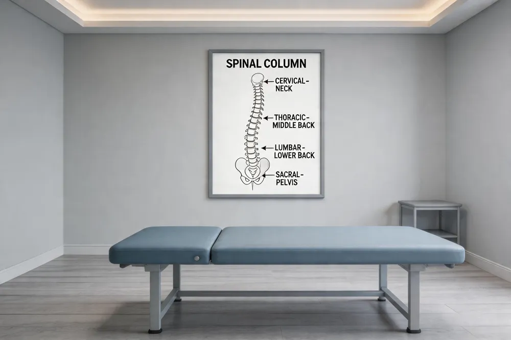
How Chiropractic Treatments Reduce Inflammation and Speed Up Back Pain Recovery

Real‑Life Stories: How Chiropractic Helped Patients Overcome Chronic Back Pain

From Symptom Masking to Root‑Cause Healing: A Practical Guide

6 Holistic Non‑Surgical Treatments That Deliver Sustainable Back‑Pain Relief

Key Questions to Ask When Selecting Your Chiropractor
Mindful Movement: Incorporating Stretch Breaks Into a Busy Day

Why Targeting the Underlying Cause Beats Temporary Symptom Relief

8 Ways Physiotherapy Complements Chiropractic Adjustments for Faster Recovery

Meal Planning for Athletes Recovering from Back Injuries

9 Everyday Lifestyle Tweaks to Preserve a Healthy Spine and Prevent Pain

Functional Movement Screening for Root‑Cause Pain Identification

6 Things You’ll Experience During Your First Chiropractic Appointment

Spinal Decompression vs. Standard Physical Therapy for Sciatica: A Comparative Review

New Findings on How Adjustments Influence Blood Circulation

Work‑From‑Home Ergonomics: A Blueprint for a Pain‑Free Back

The Link Between Chiropractic Care and Improved Sleep Quality

Physiotherapy Techniques That Strengthen the Benefits of Chiropractic Adjustments

Biomechanical Insights Behind Successful Spinal Decompression

Desk‑to‑Dumbbell: Lifestyle Shifts That Guard Your Lower Back

10 Corrective Exercises for Sustainable Pain-Free Living

Morning Routines That Support Spine Health and Reduce Stiffness

Calcium and Magnesium‑Focused Nutrition for Bone Density and Spine Health

9 Ways Chiropractic Care Transforms Back Pain Management

Testimonial: Nutrition‑Guided Recovery Accelerated by Chiropractic
Science‑Backed Spinal Decompression for Relieving Sciatica Symptoms

Key Questions to Ask During Your Initial Chiropractic Consultation
Whole‑Body Wellness Blueprint: Nutrition, Exercise, and Chiropractic Synergy

A 12‑Week Corrective Exercise Blueprint for Chronic Neck Discomfort

Manual Therapy Plus Targeted Exercise: Accelerating Post‑Injury Recovery

Home‑Based Spinal Decompression Devices: What Patients Should Know

Chiropractic Care Statistics: Reducing Lost Workdays From Back Pain

How Targeted Nutrition Enhances Immune Function and Lowers Inflammatory Pain

Self‑Assessment Tools to Identify Hidden Triggers of Back Pain

Preparing for Your First Chiropractic Appointment: A Practical Checklist

Back Pain Relief Benefits: Lower Opioid Use and Better Quality of Life

Foam‑Rolling Techniques to Complement Chiropractic Adjustments
Progressive Overload Principles in Corrective Exercise Programs

Six‑Month Transformation: Integrated Care Success Story

7 Nutritional Hacks to Boost Your Overall Wellness

How to Choose a Chiropractor: Credentials, Techniques, and Patient Compatibility

5 Ways Spinal Decompression Can Ease Sciatica Pain

Overcoming Sciatica: A Patient’s Multidisciplinary Treatment Journey
Lifestyle Changes That Promote a Healthy Spine
Physiotherapy as a Complement to Chiropractic Treatments
6 Ways Physiotherapy Enhances Chiropractic Treatment Outcomes

Daily Living Tips for a Strong and Healthy Spine

How Physiotherapy Enhances Chiropractic Outcomes
Holistic Treatments: Alternatives to Surgery for Pain Relief
Inspiring Patient Success Stories from Chiropractic Clinics
Integrating Physiotherapy with Chiropractic Treatments
Corrective Exercise Routines for Chronic Pain Prevention
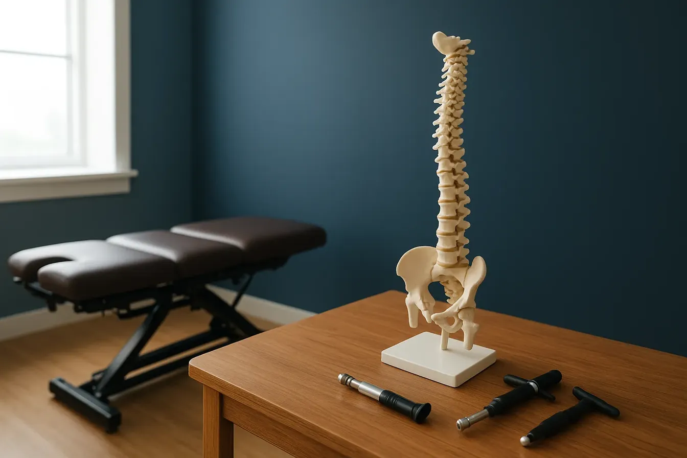
10 Corrective Exercises to Maintain a Healthy Spine and Relieve Pain
Exercise Strategies for Long-Term Pain Management
10 Corrective Exercises to Maintain a Healthy Spine and Relieve Pain
The Importance of Treating the Root Cause of Pain
Nutritional Support Strategies for Whole-Body Wellness
7 Essential Questions to Ask When Choosing Your Chiropractor
Identifying and Addressing Root Causes of Pain
Maintaining a Healthy Spine with Lifestyle Adjustments
Sciatica Symptom Relief via Spinal Decompression
8 Reasons Why Addressing the Root Cause of Pain Is Crucial
Chiropractic Care’s Impact on Back Pain Recovery

Combining Physiotherapy with Chiropractic Care for Better Results

Inspiring Recovery: Chiropractic Patient Testimonials

Nutritional Counseling Tips for Optimal Wellness

Why Addressing the Root Cause of Pain is Crucial
Back Pain and Chiropractic Care: A Winning Combination
Chiropractic Care Benefits You Might Not Know

Exploring Holistic and Non-Surgical Treatments for Pain
Spinal Health: Lifestyle Tips for Everyday Wellness
Spinal Decompression Therapy: Benefits and Applications
Holistic and Non-Invasive Spine Care Options
Everyday Lifestyle Advice to Keep Your Spine Healthy
The Significance of Treating Root Causes in Pain Therapy
Best Corrective Exercises for Sustainable Pain Management
Addressing Underlying Causes of Pain for Long-Term Relief
6 Key Benefits of Spinal Decompression Therapy for Sciatica Relief
How Nutritional Counseling Supports Spine Health and Recovery
The Role of Physiotherapy in Supporting Chiropractic Treatments